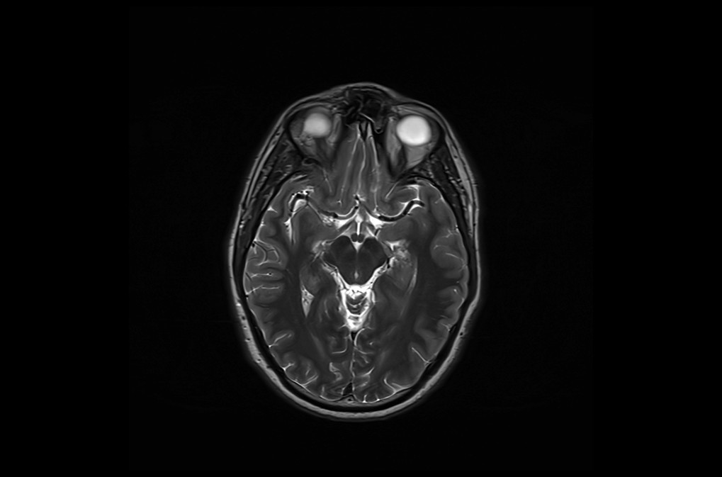
MRI Field of View (FOV)
field of view
In MRI (Magnetic Resonance Imaging), the field of view (FOV) refers to the region of the body that is imaged during the scan. It is the area of the patient’s anatomy that is captured in the image.The FOV is usually expressed in units of centimeters (cm) or millimeters (mm), and can be adjusted by the operator before the scan. A larger FOV will capture a larger area of the body, while a smaller FOV will capture a smaller area with higher spatial resolution.
The FOV is determined by the size of the imaging matrix and the physical dimensions of the MRI scanner’s gradient coils. The matrix size refers to the number of pixels in the image.
The relationship between FOV and Matrix Size can be described using the equation:
Pixel Size = FOV / Matrix Size
This equation indicates that the size of each pixel in the image (i.e., the spatial resolution) is inversely proportional to the matrix size. As the matrix size increases, the pixel size decreases, resulting in higher spatial resolution and finer detail in the image. Conversely, if the matrix size decreases, the pixel size increases, leading to lower spatial resolution and potentially more pixelation in the image
FOV 230mm

FOV 110MM

References
- Haacke, E. M., Brown, R. W., Thompson, M. R., & Venkatesan, R. (2019). Magnetic Resonance Imaging: Physical Principles and Sequence Design. John Wiley & Sons.
- Bushberg, J. T., Seibert, J. A., Leidholdt Jr, E. M., & Boone, J. M. (2011). The Essential Physics of Medical Imaging. Lippincott Williams & Wilkins.
- Stark, D. D., & Bradley, W. G. (Eds.). (2012). Magnetic Resonance Imaging. Mosby.


