MRI Epidermoid cyst
An epidermoid cyst, also known as an epidermal cyst, is a benign (non-cancerous) skin lesion that arises from the epidermis, the outermost layer of the skin. These cysts are commonly found on the face, neck, trunk, and sometimes on the genitalia, but they can occur anywhere on the skin.
Causes:
The precise cause of epidermoid cysts is not entirely understood, but they often arise from a swollen hair follicle or skin trauma. Genetics may also play a role, as these cysts sometimes appear in groups that suggest a hereditary component.
Symptoms:
Epidermoid cysts typically present as small, round bumps under the skin. These cysts may be yellow or flesh-colored and can range in size from a few millimeters to several centimeters in diameter. They grow slowly and are typically not painful. However, symptoms may include:
- A noticeable lump on the skin
- A blackhead at the center of the cyst
- Redness or swelling if inflamed or infected
- Soreness or discomfort if the cyst is located in an area that is frequently irritated
- A cheesy or oily material that can drain from the cyst
Treatment:
Treatment for epidermoid cysts usually depends on whether they are causing discomfort or cosmetic concerns. Options include:
Minor surgery: This is the most definitive treatment and involves removing the entire cyst to prevent recurrence. It’s usually done under local anesthesia
Incision and drainage: This provides temporary relief from symptoms but does not remove the cyst wall, so the cyst may recur.
Injections: Steroid injections can reduce inflammation if the cyst is swollen.
Antibiotics: If an epidermoid cyst becomes infected, antibiotics might be prescribed to clear the infection.
MRI Appearance of Epidermoid cyst
T1-Weighted (T1W) Images:
Epidermoid cysts typically appear as well-defined lesions with low to intermediate signal intensity on T1W images.
T2-Weighted (T2W) Images:
On T2W images, these cysts often show high signal intensity, similar to cerebrospinal fluid (CSF). They are typically brighter than the surrounding brain parenchyma due to their high water content.
Short Tau Inversion Recovery (STIR):
Epidermoid cysts are usually hyperintense on STIR sequences. This sequence is helpful in suppressing fat signal and highlighting the cyst against the surrounding tissue, which may be useful in detecting inflammation or edema.
Diffusion-Weighted Imaging (DWI):
Epidermoid cysts characteristically appear very bright on DWI, and this is one of the key sequences for their diagnosis. They demonstrate restricted diffusion because the keratin content and the lamellated structure of the cysts impede the movement of water molecules.
Post-Contrast T1 with Fat Saturation (Fat-Sat):
After the administration of gadolinium contrast, there is usually no significant enhancement within the cyst itself, given its avascular nature. However, any associated inflammation or infection may show peripheral enhancement. The fat-saturation technique can help to suppress the signal from surrounding fat, making any enhancement more conspicuous.
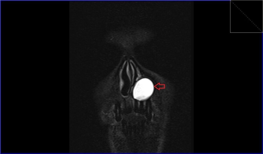
STIR coronal image shows Epidermoid cyst
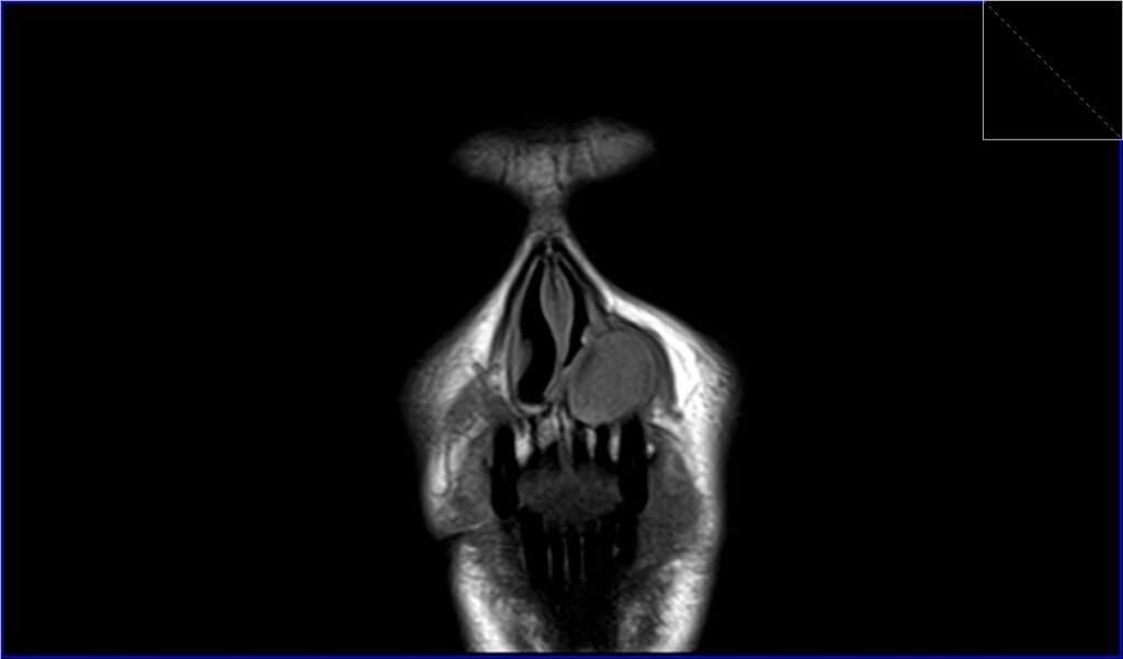
T1 coronal image shows Epidermoid cyst
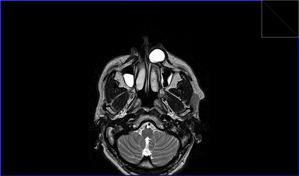
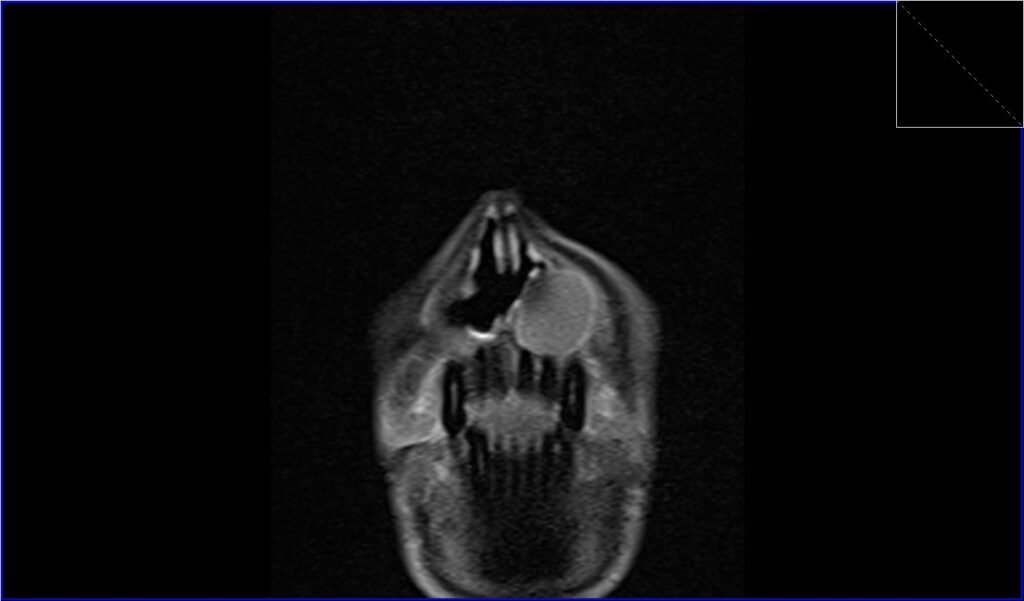
T2 axial image shows Epidermoid cyst
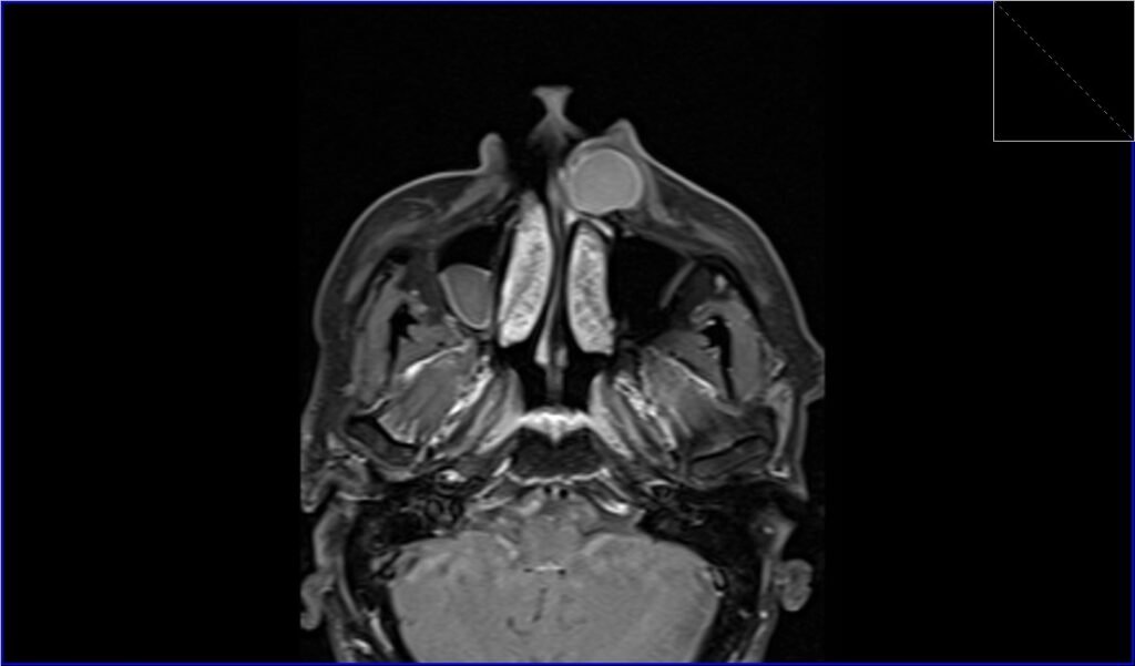
T1 fat sat post contrast axial image shows Epidermoid cyst
T1 fat sat post contrast coronal image shows Epidermoid cyst
References
- Hong, S. H., Chung, H. W., Choi, J. Y., Koh, Y. H., Choi, J. A., & Kang, H. S. (2006). MRI Findings of Subcutaneous Epidermal Cysts: Emphasis on the Presence of Rupture. American Journal of Roentgenology, 186(4).
- Hoang VT, Trinh CT, Nguyen CH, Chansomphou V, Chansomphou V, Tran TTT. Overview of epidermoid cyst. Eur J Radiol Open. 2019;6:291–301. doi: 10.1016/j.ejro.2019.08.003. PMCID: PMC6732711. PMID: 31516916
- Law, E. K. C., Lee, R. K. L., Ng, A. W. H., Siu, D. Y. W., & Ng, H.-K. (2015). Atypical Intracranial Epidermoid Cysts: Rare Anomalies with Unique Radiological Features. Case Report, Volume 2015, Article ID 528632.
- Lim, J., & Cho, K. (2015). Epidermoid cyst with unusual magnetic resonance characteristics and spinal extension. World Journal of Surgical Oncology, 13, 240. doi:10.1186/s12957-015-0629-4
- Hong, S. H., Chung, H. W., Choi, J.-Y., Koh, Y. H., Choi, J.-A., & Kang, H. S. (2006). MRI Findings of Subcutaneous Epidermal Cysts: Emphasis on the Presence of Rupture. Musculoskeletal Imaging • Clinical Observations, 186, 961–966. doi:10.2214/AJR.05.0044.
- Phillips, J., & Chiu, L. (1987). Magnetic resonance imaging of intraspinal epidermoid cyst: A case report. Journal of Computed Tomography, 11(2), 181-183.