Humerus Enchondroma MRI
An enchondroma is a benign (non-cancerous) tumor that forms in the cartilage within the bones. These tumors most commonly occur in the small bones of the hands and feet, but they can also develop in other bones such as the femur, humerus, and tibia. Enchondromas are typically found incidentally during imaging for other conditions, as they often do not cause symptoms.
Causes
The exact cause of enchondromas is not well understood. However, it is believed to be associated with abnormal growth and development of cartilage cells. There is no clear link to genetic factors or lifestyle influences, and they occur sporadically.
Symptoms
Most enchondromas do not cause any symptoms and are found incidentally. However, if symptoms do occur, they may include:
- Pain: If the tumor grows and exerts pressure on surrounding tissues or nerves.
- Swelling: In the affected area.
- Bone fractures: Weakened bones due to the presence of the tumor may fracture easily.
- Deformity: In rare cases, especially if the tumor is large or located in a critical area.
Diagnosis
Diagnosis of an enchondroma typically involves:
- Medical History and Physical Examination: The doctor will review the patient’s medical history and conduct a physical examination to check for signs and symptoms.
- Imaging Studies:
- X-rays: Initial imaging to identify bone abnormalities.
- MRI (Magnetic Resonance Imaging): Provides detailed images of the bone and surrounding tissues to assess the size and extent of the tumor.
- CT Scan (Computed Tomography): Sometimes used for detailed cross-sectional images.
Treatment
Treatment depends on the size and location of the enchondroma, as well as whether it is causing symptoms:
- Observation: If the enchondroma is asymptomatic, regular monitoring with periodic X-rays may be recommended to ensure it is not growing or causing problems.
- Surgical Removal: If the enchondroma is causing symptoms such as pain, swelling, or fractures, surgical removal may be necessary. Surgery may involve curettage (scraping out the tumor) and filling the cavity with bone graft material.
MRI Appearance of Enchondroma
T1 Appearance
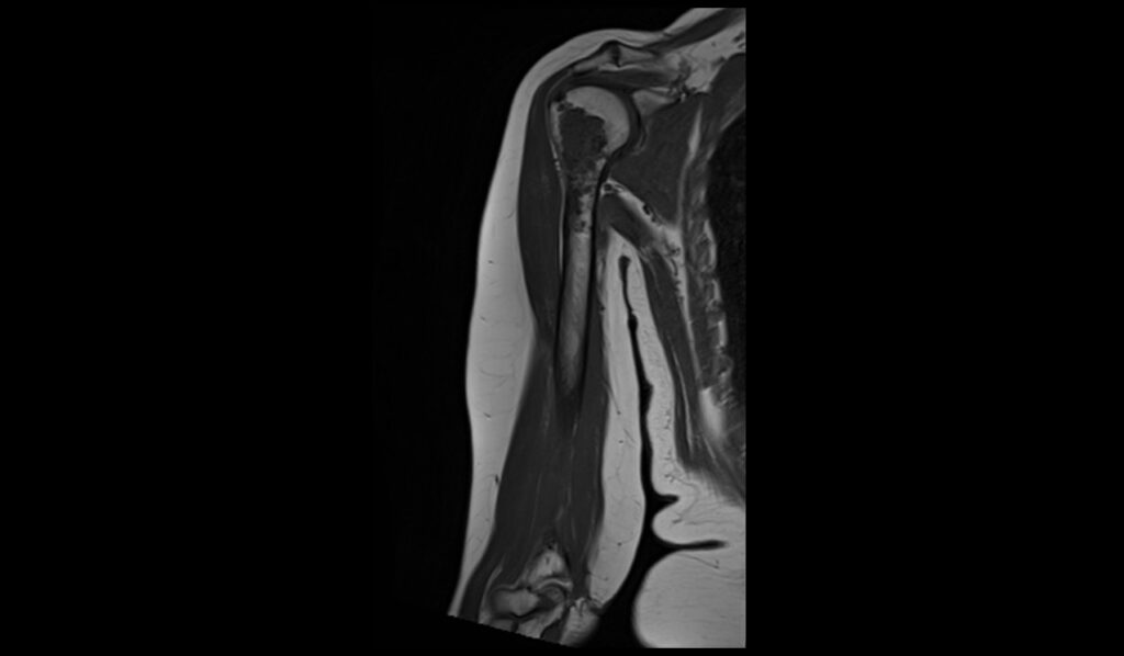
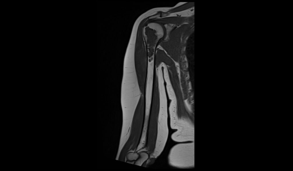
On T1-weighted MRI images, enchondromas typically present as lesions with low to intermediate signal intensity relative to normal bone marrow. The internal structure of these benign cartilaginous tumors may show heterogeneous signal due to the presence of calcifications and cartilaginous matrix. Sometimes, regions of higher signal intensity may be observed within the lesion, reflecting areas of marrow fat or myxoid change within the tumor matrix. These characteristics help in distinguishing enchondromas from other bone lesions.
T2 Appearance
Enchondromas are more conspicuous on T2-weighted MRI images, where they usually display high signal intensity due to their high water content within the hyaline cartilage matrix. The lesion may appear even more heterogeneous on T2 sequences due to the presence of calcifications, which are hypointense. The high contrast between the hyperintense cartilaginous areas and hypointense calcifications often aids in the diagnosis of enchondroma.
STIR Appearance
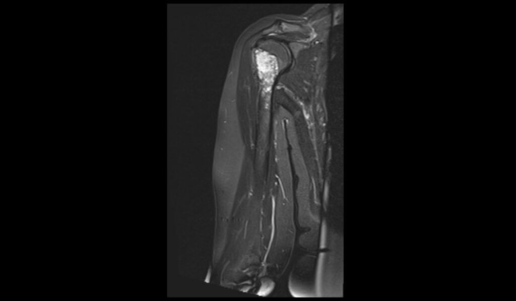
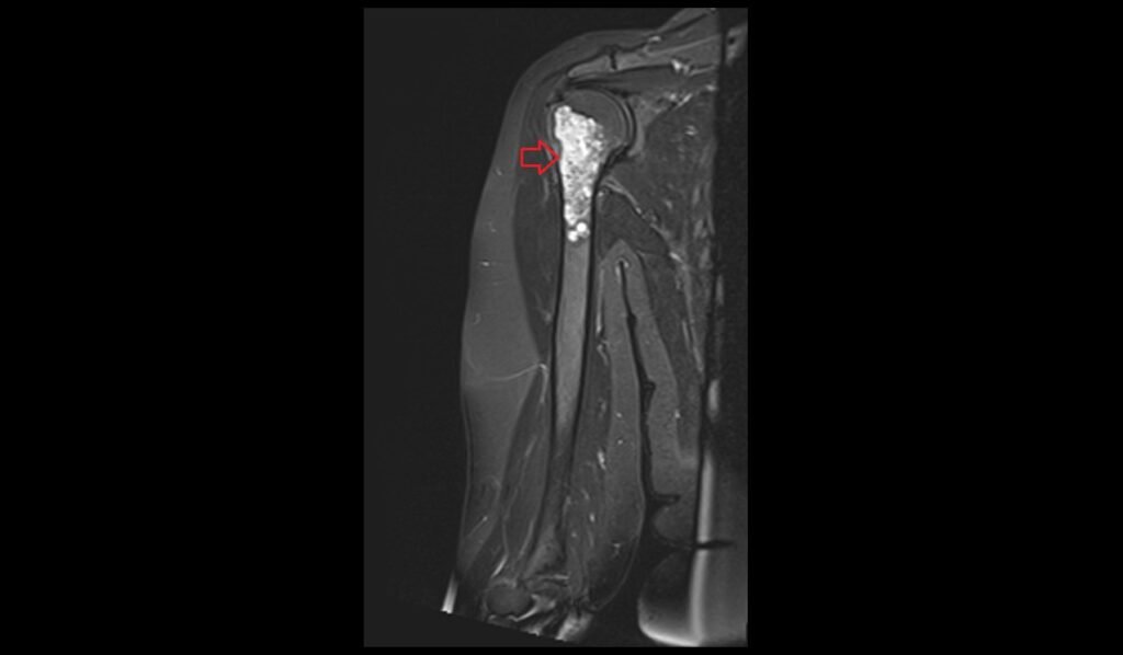
On Short Tau Inversion Recovery (STIR) images, enchondromas show a marked hyperintense signal, which effectively suppresses the signal from surrounding fat, enhancing the visibility of the lesion. This high signal intensity is characteristic of the water-rich cartilaginous matrix of the tumor. The use of STIR imaging is particularly useful for detecting subtle lesions within the bone and for evaluating the extent of marrow involvement, providing valuable information that complements findings from other MRI sequences.
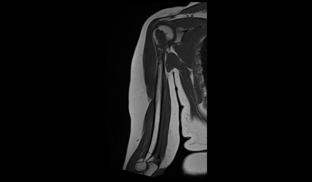
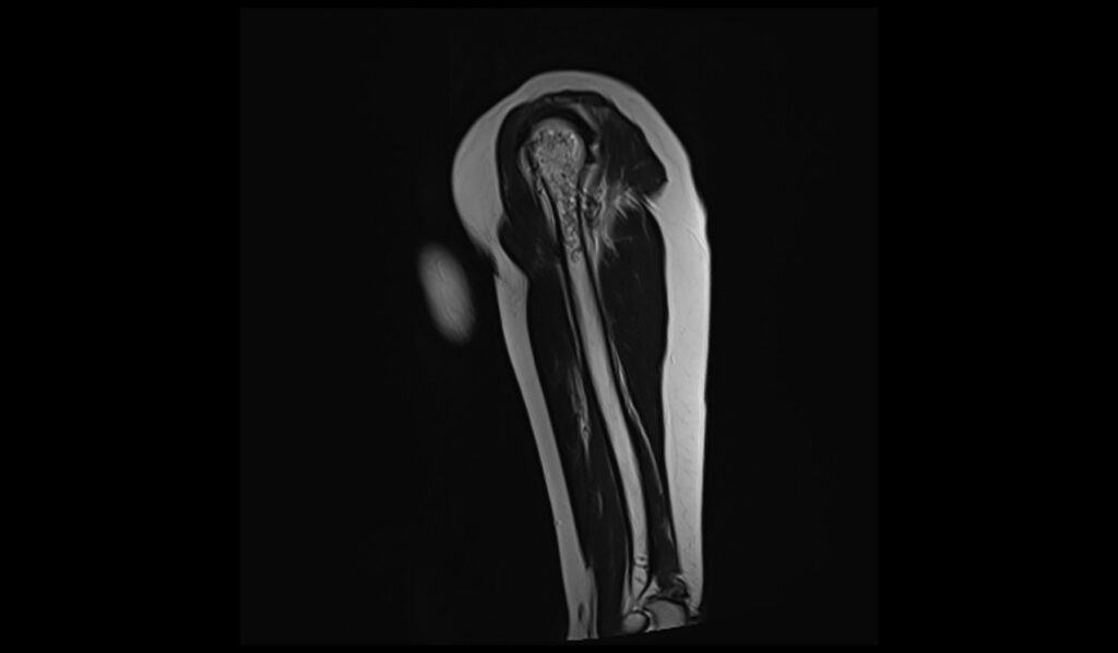
STIR coronal image shows Humerus Enchondroma

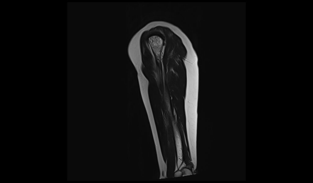
T1 TSE coronal image shows Humerus Enchondroma
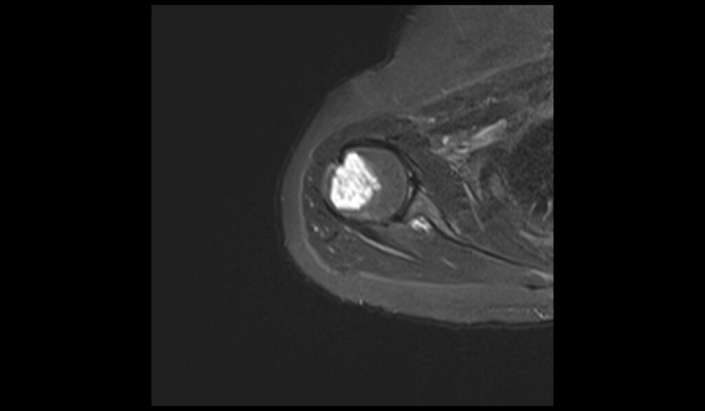
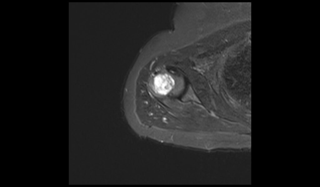
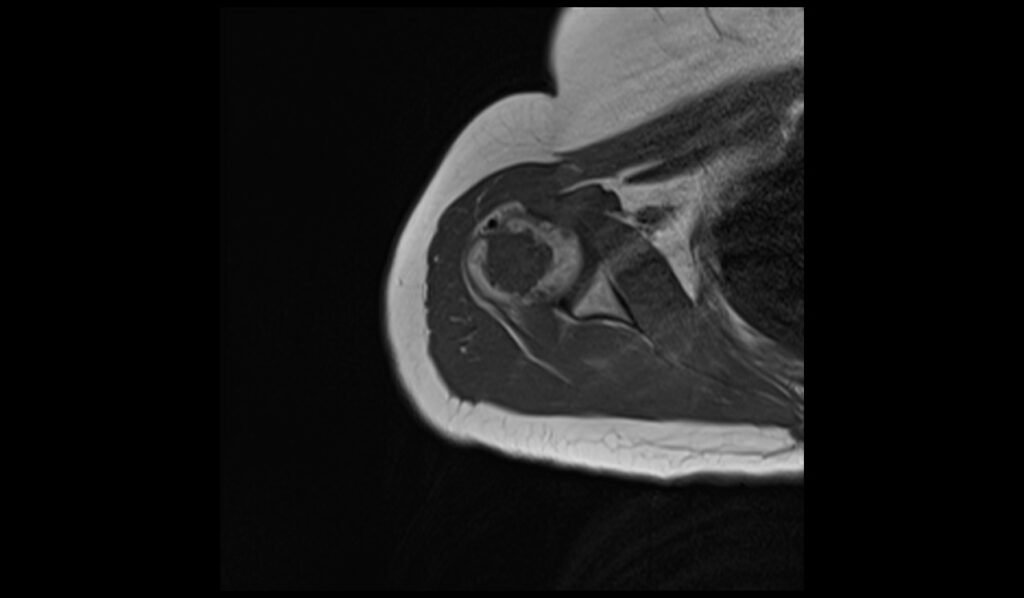
STIR axial image shows Humerus Enchondroma

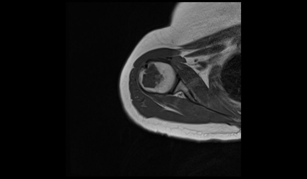
T1 axial image shows Humerus Enchondroma
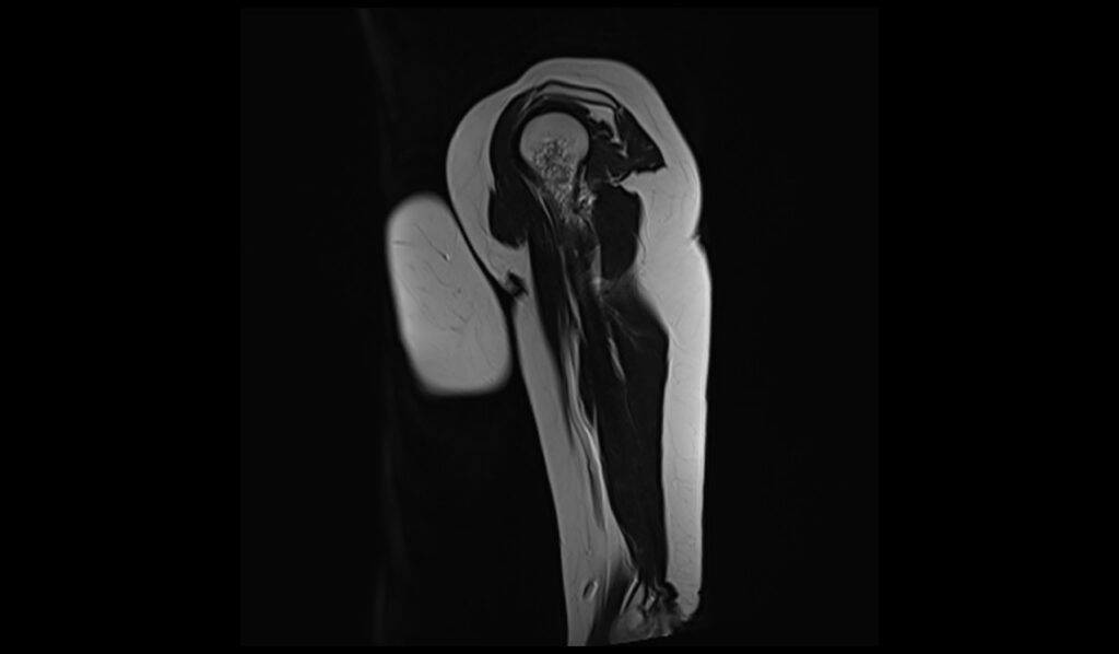
T2 sagittal image shows Humerus Enchondroma
References
- Omlor, G. W., Lohnherr, V., Lange, J., Gantz, S., Merle, C., Fellenberg, J., Raiss, P., & Lehner, B. (2018). Enchondromas and atypical cartilaginous tumors at the proximal humerus treated with intralesional resection and bone cement filling with or without osteosynthesis: Retrospective analysis of 42 cases with 6 years mean follow-up. World Journal of Surgical Oncology, 16, Article 139. https://doi.org/10.1186/s12957-018-1441-3
- Walden, M. J., Murphey, M. D., & Vidal, J. A. (2008). Incidental enchondromas of the knee. American Journal of Roentgenology, 190(6). https://doi.org/10.2214/AJR.07.2796
- Levy, J. C., Temple, H. T., Mollabashy, A., Sanders, J., & Kransdorf, M. (2005). The causes of pain in benign solitary enchondromas of the proximal humerus. Clinical Orthopaedics and Related Research, 431, 181-186. https://doi.org/10.1097/01.blo.0000150114.19489.c4