MRI cerebrospinal fluid (CSF) leak
A cerebrospinal fluid (CSF) leak occurs when the fluid that surrounds and cushions the brain and spinal cord leaks through a tear or hole in the dura mater, the outermost layer of the meninges.
Causes
- Trauma or Injury: Physical trauma, such as a fall or an accident, can cause a tear in the dura mater.
- Surgery: Spinal or cranial surgeries can sometimes result in a CSF leak.
- Spontaneous Leak: This can occur without any obvious cause, often related to connective tissue disorders like Marfan syndrome or Ehlers-Danlos syndrome.
- Epidural Injections or Spinal Taps: Medical procedures that involve inserting needles into the spine can sometimes cause CSF leaks.
Symptoms
- Headache: A positional headache that worsens when upright and improves when lying down is a hallmark symptom.
- Neck and Shoulder Pain: Pain or stiffness in the neck and shoulders.
- Nausea and Vomiting: Often associated with the headache.
- Visual Disturbances: Blurred or double vision, sensitivity to light.
- Tinnitus: Ringing in the ears.
- Hearing Changes: Hearing loss or a sensation of fullness in the ears.
- Cranial Nerve Palsies: Weakness or paralysis of the muscles controlled by the cranial nerves.
- Dizziness or Vertigo: A sensation of spinning or imbalance.
- Cognitive Changes: Memory issues, difficulty concentrating.
Diagnosis
- MRI and CT Scans: Imaging techniques such as MRI (especially with T1, T2, T2 3D with high TE and STIR sequences) and CT myelography can help visualize the leak and its location.
- Lumbar Puncture: This procedure can measure the pressure of the CSF and collect samples for analysis.
Treatment
- Conservative Management: Bed rest, hydration, and caffeine intake to help reduce symptoms.
- Epidural Blood Patch: Injection of the patient’s blood into the epidural space to create a clot and seal the leak.
Surgery: In cases where conservative treatment fails or the leak is severe, surgical repair of the dura mater may be necessary.
Fibrin Glue Injection: Sealing the leak with a special adhesive.
MRI Appearance of CSF Leak
T1-Weighted Imaging (T1WI)
- Appearance: CSF is typically hypointense (dark) on T1-weighted images.
- CSF Leak: The leak may appear as a hypointense signal along the site of the dural tear. Adjacent tissues may show signs of edema or other abnormalities.
T2-Weighted Imaging (T2WI)
- Appearance: CSF is hyperintense (bright) on T2-weighted images.
- CSF Leak: The leak is visualized as a hyperintense signal at the site of the dural defect. The surrounding tissues might also show hyperintensity due to the presence of CSF.
Short Tau Inversion Recovery (STIR)
- Appearance: This sequence suppresses fat signals and highlights fluid, making CSF appear bright.
- CSF Leak: The leak appears as a hyperintense signal against the suppressed background. STIR sequences are particularly useful for identifying fluid collections and differentiating them from fat.
T2 SPACE 3D with High TE (Echo Time)
- Appearance: This is a heavily T2-weighted sequence providing high-resolution, three-dimensional images with excellent contrast between fluid and other tissues.
- CSF Leak: T2 SPACE 3D sequences are highly sensitive in detecting CSF leaks. The high echo time enhances the signal from fluid, making the CSF leak and any associated meningeal diverticula or pseudomeningoceles stand out as bright areas. This sequence provides detailed anatomical information, crucial for surgical planning if needed.
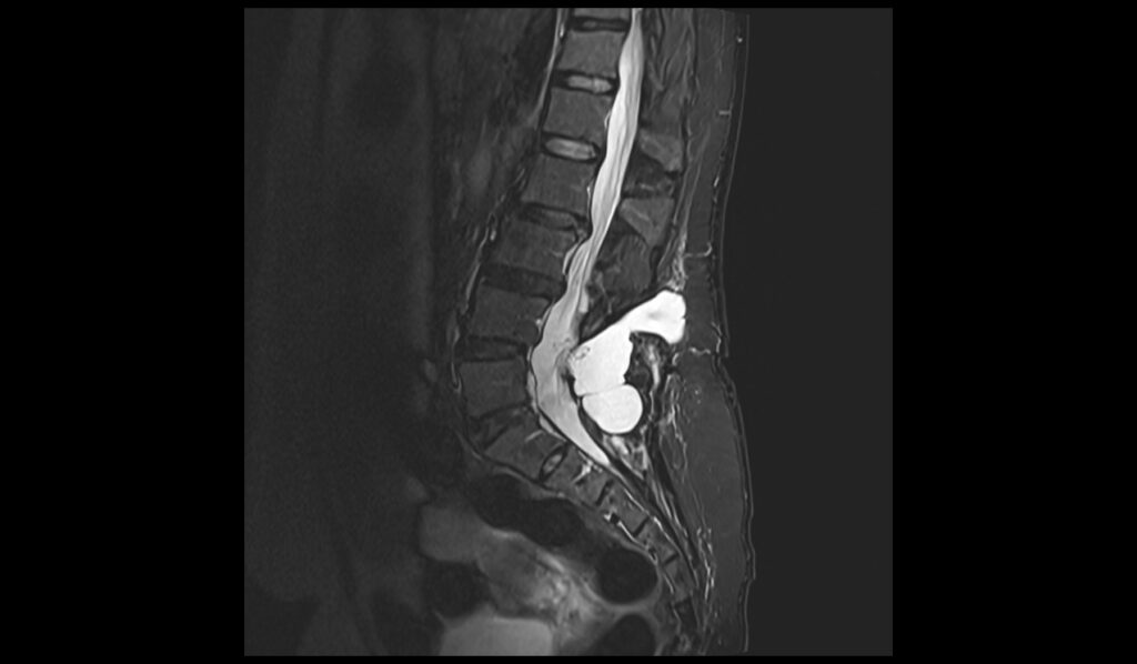
T2 sagittal image of lumbar spine shows CSF Leak

STIR sagittal image of lumbar spine shows CSF Leak
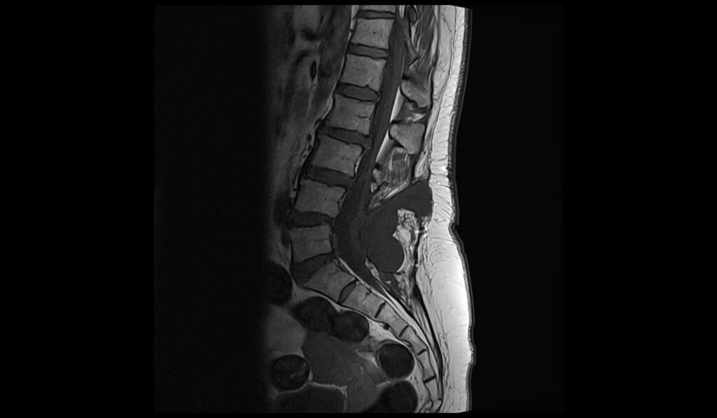
T1 TSE sagittal image of lumbar spine shows CSF Leak
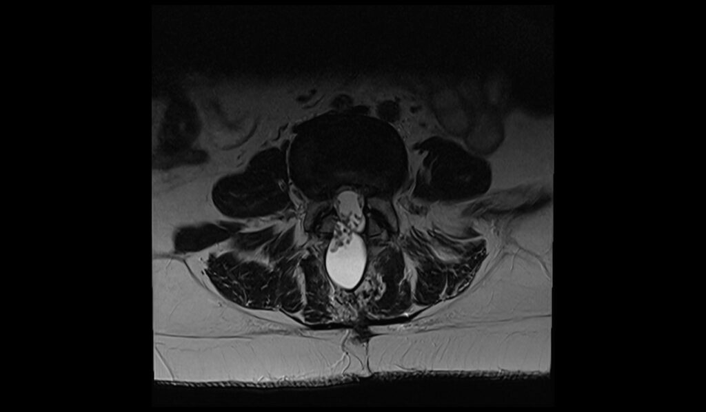
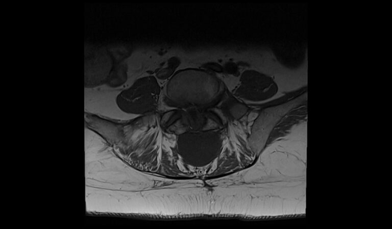
T2 TSE axial image of lumbar spine shows CSF Leak
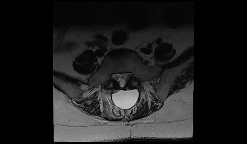
T1 TSE axial image of lumbar spine shows CSF Leak

References
- Medina, J. H., Abrams, K., Falcone, S., & Bhatia, R. G. (2010). Spinal imaging findings in spontaneous intracranial hypotension. AJR American Journal of Roentgenology, 195(2), 459-464.
- Farb, R.I., Nicholson, P.J., Peng, P.W., Massicotte, E.M., Lay, C., Krings, T., & terBrugge, K.G. (2019). Spontaneous intracranial hypotension: A systematic imaging approach for CSF leak localization and management based on MRI and digital subtraction myelography. American Journal of Neuroradiology, 40(4), 745-753.
- Dobrocky T, Grunder L, Breiding PS, et al. Assessing Spinal Cerebrospinal Fluid Leaks in Spontaneous Intracranial Hypotension With a Scoring System Based on Brain Magnetic Resonance Imaging Findings. JAMA Neurology. 2019;76(5):580-587.
- Akiba, C., Bandai, H., Ito, Y., Maeda, T., Yamaguchi, K., Nakajima, M., & Miyajima, M. (2020). Cerebrospinal fluid leak presented with the C1-C2 sign caused by spinal canal stenosis: a case report. BMC Neurology, 20(151).