Choroid Plexus Papilloma MRI
Choroid plexus papilloma is a rare, benign tumor of the choroid plexus, which is a network of cells that produce cerebrospinal fluid (CSF) in the brain. Although benign, Choroid plexus papilloma can lead to significant health issues due to increased CSF production and subsequent hydrocephalus.
Causes
The exact cause of choroid plexus papilloma is not well understood. However, like many tumors, it is believed to be related to genetic mutations that lead to uncontrolled cell growth. These tumors are generally sporadic, meaning they do not typically run in families.
Symptoms
Symptoms of choroid plexus papilloma can vary depending on the size and location of the tumor but are generally related to increased intracranial pressure due to excess CSF production. Common symptoms include:
- Headaches
- Nausea and vomiting
- Enlarged head size in infants (macrocephaly)
- Hydrocephalus (accumulation of CSF in the brain)
- Irritability in infants and young children
- Vision problems
- Difficulty with balance and coordination
- Seizures
Diagnosis
Diagnosing CPP involves a combination of clinical evaluation and imaging techniques:
- Clinical Evaluation: Assessment of symptoms and physical examination.
- MRI and CT Scans: These imaging tools are essential for visualizing the location, size, and effect of the tumor on surrounding structures. MRI is particularly useful for detailed imaging of brain tissue.
- Biopsy: A biopsy may be performed during surgery to confirm the diagnosis by examining the tissue under a microscope.
Treatment
Treatment for choroid plexus papilloma typically involves a combination of the following approaches:
- Surgical Removal: The primary treatment is complete surgical resection of the tumor. This can often result in a cure, especially if the tumor is benign and fully removed.
- Management of Hydrocephalus: If hydrocephalus is present, additional procedures, such as the placement of a shunt, may be needed to drain excess CSF and relieve intracranial pressure.
- Follow-up Care: Regular follow-up with imaging studies to monitor for recurrence of the tumor.
MRI appearance of Choroid Plexus Papilloma
MRI T1 Appearance of Choroid Plexus Papilloma
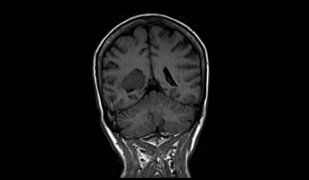
Choroid plexus papilloma typically presents as iso- to hypo-intense on T1-weighted MRI images. This characteristic appearance helps in differentiating it from other intracranial lesions, as the relative intensity on T1 helps to highlight the nature of the tissue compared to surrounding cerebral structures, emphasizing the solid component of the tumor.
MRI T2 Appearance of Choroid Plexus Papilloma
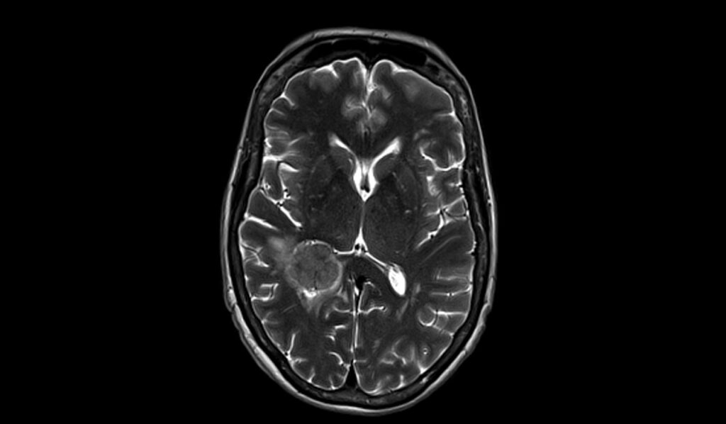
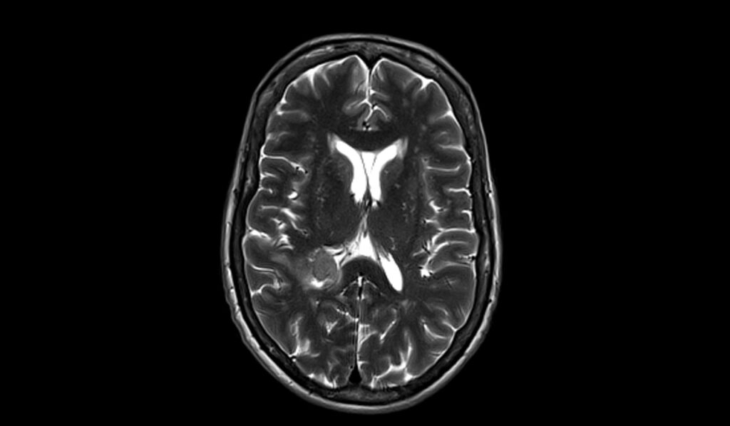
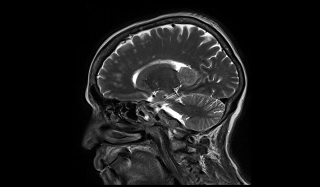
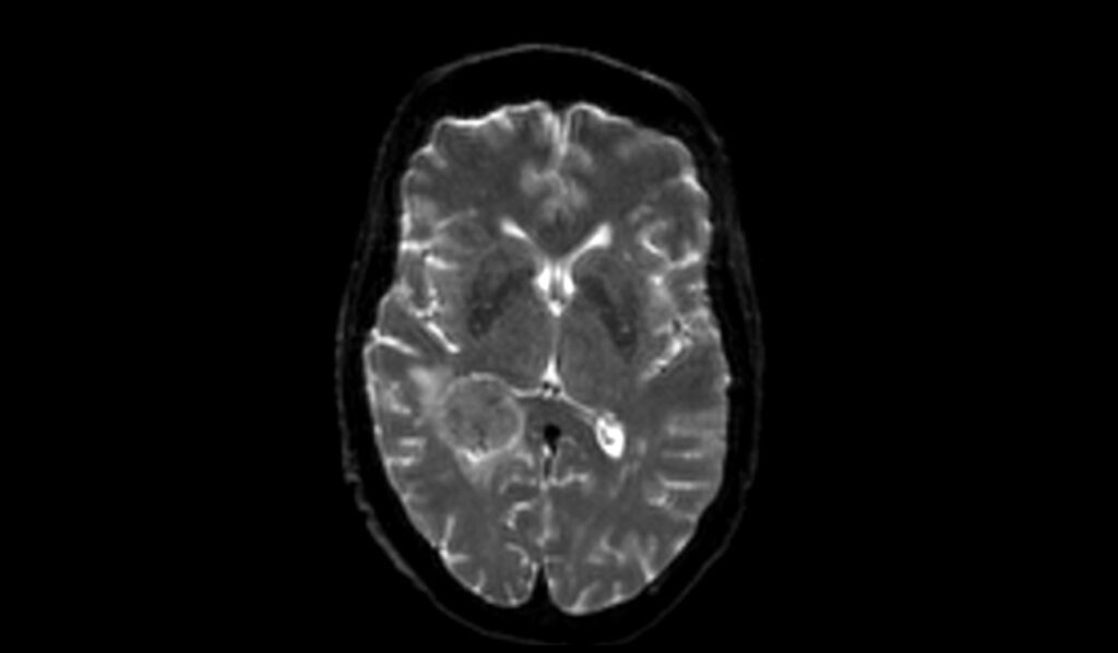
On T2-weighted MRI scans, choroid plexus papillomas are typically bright, indicating high water content within the tumor. This brightness stands out against the generally darker appearance of the surrounding brain tissue and ventricles, aiding in the precise localization and assessment of the extent of the tumor.
MRI FLAIR Appearance of Choroid Plexus Papilloma
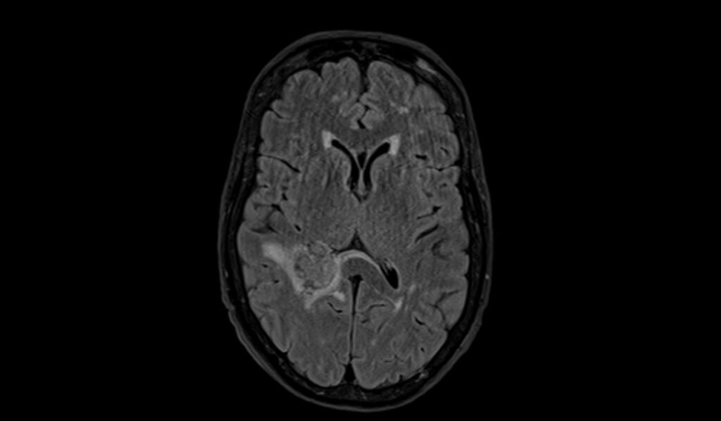
FLAIR (Fluid Attenuated Inversion Recovery) imaging of choroid plexus papillomas shows a bright signal. This modality is particularly useful for suppressing the cerebrospinal fluid (CSF) signal, thereby enhancing the contrast between the lesion and the CSF, which is particularly valuable in lesions adjacent to CSF spaces.
MRI DWI (b0, b1000) and ADC Appearance of Choroid Plexus Papilloma
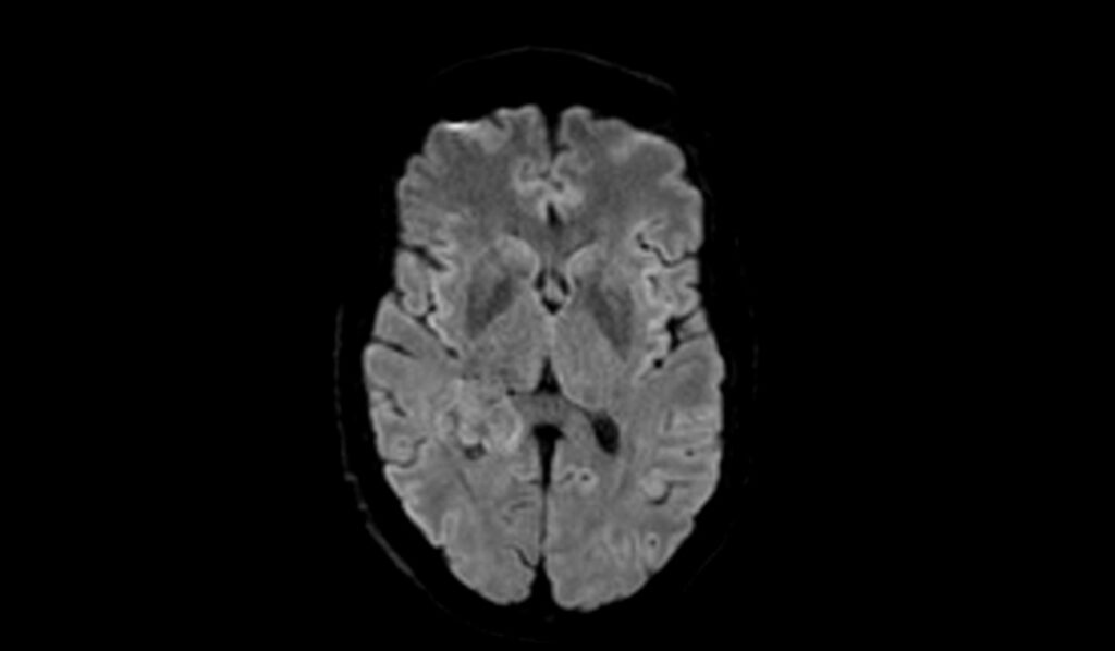
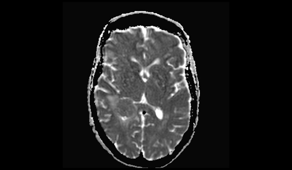
Diffusion Weighted Imaging (DWI) reveals that choroid plexus papillomas are typically bright on b0 images, indicating unrestricted diffusion in its free water molecules. Conversely, on b1000 images, the lesion appears hyperintense, suggesting restricted diffusion within the tumor cells. The apparent diffusion coefficient (ADC) maps usually show these tumors as dark, indicating a lower ADC value typical of cellular tumors, which helps in distinguishing them from cystic or necrotic lesions.
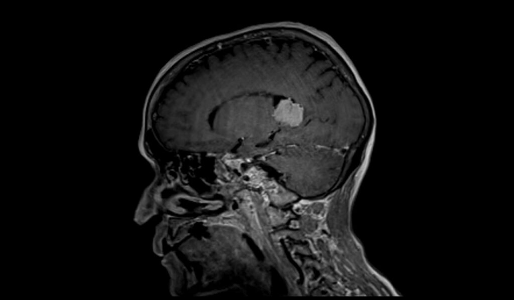
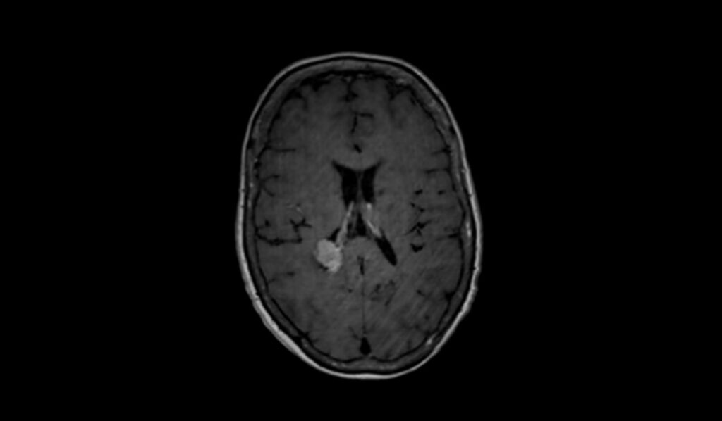
MRI T1 Post-Contrast Appearance of Choroid Plexus Papilloma
After the administration of contrast material, choroid plexus papillomas typically exhibit very bright enhancement on T1-weighted images. This marked enhancement is due to the vascular nature of the tumor and significantly aids in delineating the tumor from adjacent brain tissues, enhancing diagnostic accuracy and surgical planning.
T2 axial images shows Choroid Plexus Papilloma

FLAIR axial image shows Choroid Plexus Papilloma
T1 coronal image shows Choroid Plexus Papilloma
T2 sagittal image shows Choroid Plexus Papilloma
DWI b0 image shows Choroid Plexus Papilloma
DWI b1000 image shows Choroid Plexus Papilloma
DWI ADC map image shows Choroid Plexus Papilloma
T1 sagittal post contrast images shows Choroid Plexus Papilloma


T1 coronal post contrast image shows Choroid Plexus Papilloma

T1 axial post contrast images shows Choroid Plexus Papilloma

References
- Andour, H., Ben El Hend, S., Mandour, C., Allaoui, M., & Fikri, A. (2024). Atypical choroid plexus papilloma: Diagnosis and differentials: A case report. SAGE Open Medical Case Reports, 12, 2050313X241254000.
- Lin, H., Leng, X., Qin, C., Du, Y., Wang, W., & Qiu, S. (2019). Choroid plexus tumours on MRI: similarities and distinctions in different grades. Cancer Imaging, 19(1), 17. https://doi.org/10.1186/s40644-019-0200-1
- Muñoz Montoya, J. E., Maldonado Moran, M. A., Santamaria Rodriguez, P., Toro Lopez, S., Perez Cataño, C. S., & Luque Suarez, J. C. (2019). Choroid plexus papilloma of the fourth ventricle: A pediatric patient. Asian Journal of Neurosurgery, 14(2), 585-588.
- Sethi, D., Arora, R., Garg, K., & Tanwar, P. (2017). Choroid plexus papilloma. Asian Journal of Neurosurgery, 12(1), 139-141. https://doi.org/10.4103/1793-5482.153501