SPACE/CUBE/VISTA
SPACE (Sampling Perfection with Application optimized Contrasts using different flip-angle Evolutions) is a 3D TSE (turbo spin echo) sequence used in magnetic resonance imaging (MRI). It’s known for providing high-resolution isotropic 3D images, which can be reformatted in any plane without loss of image quality. This makes the SPACE sequence particularly useful for imaging structures with complex anatomy or for cases where multiplanar reconstructions are needed.
SPACE is a variant of the traditional three-dimensional (3D) turbo spin echo sequence used in MRI. Differing from the conventional method, SPACE employs non-selective, brief refocusing pulse trains made up of RF pulses with changing flip angles. This novel approach facilitates very high turbo factors exceeding 100, leading to enhanced sampling efficiency.
One of the remarkable features of SPACE is its capability to yield high-resolution isotropic images, which can then be post-processed and visualized in multiple orientations. This adaptability offers an edge over other techniques. Moreover, SPACE sequences showcase a higher resistance to common MRI challenges like susceptibility, flow, and chemical shift artifacts, ensuring clearer and more accurate imaging. This resistance gives SPACE a distinct advantage over the traditional turbo spin echo sequences.
One of the practical benefits of the SPACE technique is its speed and efficiency. Leveraging this sequence, it becomes feasible to produce isotropic 0.9mm 3D images with T1,T2,PD, FLAIR, and STIR contrasts in a time frame of less than 6 minutes, making it an invaluable tool in advanced diagnostic imaging.
SPACE MRI Applications
Neuroimaging:
- Cranial Nerve Imaging: SPACE provides high-resolution images, particularly useful for delineating cranial nerve anatomy, especially those in close proximity to bone or air, such as the trigeminal nerve or vestibulocochlear nerve.
- FLAIR 3D for MS Imaging: Multiple sclerosis (MS) lesions, especially those in the periventricular region or spinal cord, can be visualized in detail using the FLAIR (Fluid-attenuated inversion recovery) 3D SPACE sequence. The isotropic nature of this sequence allows for multiplanar reconstructions, making lesion identification and volume calculations easier and more accurate.
- T1 Volumetric 3D for Brain Imaging: This sequence is optimized for T1-weighted brain imaging. It is useful for evaluating various neurological conditions, characterizing lesions, and performing volumetric studies of brain structures.
Abdominal Imaging:
- MRCP: SPACE sequences, especially heavily T2-weighted variants, are ideal for MR Cholangiopancreatography (MRCP) studies. They can clearly delineate the biliary tree and pancreatic ducts without the need for contrast, providing a non-invasive alternative to procedures like ERCP.
Musculoskeletal Imaging:
- MSK 3D: This sequence is optimized for imaging small structures such as tendons or cartilage. SPACE’s high resolution and contrast capabilities make it valuable for visualizing fine anatomical details, including small tears or degenerative changes.
Vascular Imaging:
SPACE Vessel Sequence: This sequence includes both dark-blood and bright-blood techniques.
Dark-Blood Imaging: SPACE can be tailored for “dark-blood” imaging, effectively suppressing signals from flowing blood and highlighting vascular pathologies.
Bright-Blood Imaging with NATIVE SPACE Vessel Sequence: This modification of the SPACE sequence for vascular imaging without contrast provides detailed images of the vessel lumen. It is particularly valuable for identifying aneurysms, stenoses, or other vascular abnormalities.
Use of SPACE MRI Sequence
- Very useful for 3D MRCP imaging (respiratory gated SPACE scans)
- Very useful for brain imaging (T1,T2 and FLAIR 3D)
- Very useful for musculoskeletal imaging ( e.g. 3D fat sat SPACE knee)
- Very useful for spinal cord imaging
- Very useful for cranial nerves imaging (e.g. IAMs and trigeminal nerve)
- Very useful for face imaging (T1 and 3D SPACE STIR in orbits, sinuses and cheek)
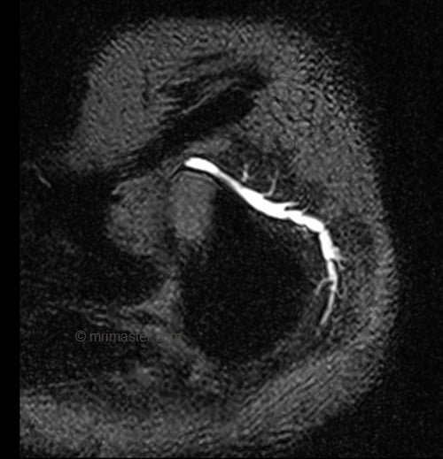
- Very useful for sialography imaging (.6MM 3D T2 SPACE with high TE)
- Useful for neck imaging
T2 SPACE axial sequence used in iam's imaging

T2 SPACE sagittal sequence used in sialography imaging

T2 SPACE fat sat coronal sequence used in MRCP imaging

T1 SPACE 3D Sagittal sequence Used in brain imaging


