Short Tau Inversion Recovery (STIR) MRI
STIR (Short Tau Inversion Recovery) imaging is a specialized MRI technique designed to enhance the visibility of abnormalities in tissues by suppressing the signal from fat. Through the strategic use of inversion pulses and short inversion times, STIR imaging creates high contrast between fat and other structures, making fluid-filled regions, edema, and inflammation stand out more prominently. This technique is especially valuable in musculoskeletal imaging, where it aids in diagnosing fractures, bone marrow disorders, and joint conditions.
STIR MRI Sequence Physics
The inversion recovery sequences are spin echo sequences with a 180° preparation pulse that flips the longitudinal magnetization in the opposite direction (i.e., spins are flipped to the 180° position). Transverse magnetization remains at zero; therefore, we do not receive an MR signal. During the recovery period, the negative longitudinal magnetization decays to zero and then begins to rise. Since transverse magnetization is not achievable, no signal is measured. To generate an MR signal, the longitudinal magnetization is then converted to transverse magnetization through the application of a 90° pulse. The time interval between the 180° pulse and the 90° excitation pulse is known as the inversion time (TI). As the spins relax back to their equilibrium configuration, the signal for each spin group evolves from a negative signal, which is zero at the null point, to a positive signal. This rate of evolution is determined by the T1 of the spin group. For instance, at 1.5 T, T1 for fat is approximately 260 ms, while for most other tissues, T1 is greater than 500 ms. Therefore, the null point for the fat signal occurs sooner than for other tissues. As a result, an inversion recovery sequence with a short inversion time (TI) of 130-150 ms is used for fat suppression.

MRI image appearance of STIR images
The easiest way to identify STIR images is to look for fat- and fluid-filled spaces in the body (e.g. cerebrospinal fluid in the brain ventricles and spinal canal, free fluid in the abdomen, fluid in the gall bladder and common bile duct, synovial fluid in joints, fluid in the urinary tract and urinary bladder, edema or any other pathological fluid collection in the body). Fluids normally appear bright, and fat appears very dark in a STIR image.
Tissues and their STIR appearance
Brain:
- CSF: Bright.
- Fat: Dark.
- White Matter: Intermediate to dark.
- Gray Matter: Intermediate to bright.
- Bone (skull): Dark.
- Bone Marrow: Intermediate to dark.
- Blood Vessels: Mostly Dark; Depending on flow characteristics, can be bright or dark
- Pituitary Gland: Intermediate.
- Choroid Plexus: Intermediate.
- Cerebellum: Gray matter Brighter than white matter.
- Brain Stem: Intermediate.
- Sinuses: Dark (air-filled).
- Thalamus, Putamen, Hippocampus, Caudate Nucleus: Intermediate.
- Corpus Callosum: Intermediate.
- Pineal Gland: Intermediate.
Spine:
- Spinal Cord: Intermediate.
- CSF: Bright.
- Bone: Dark.
- Bone Marrow: Intermediate to dark.
- Intervertebral Disc: Intermediate to bright
- Ligaments: Intermediate to Dark.
- Nerve Roots: Intermediate.
Abdomen and Pelvis:
- Liver: Intermediate to dark.
- Gallbladder and Common Bile Duct: Bright
- Spleen: Intermediate to dark.
- Kidney: Intermediate.
- Ureters: Intermediate.
- Pancreas: Intermediate to dark.
- Urinary Bladder: Bright.
- Prostate: Intermediate.
- Uterus: Intermediate.
Musculoskeletal:
- Muscle: Intermediate to dark
- Bone: Dark (low signal)
- Bone Marrow: Intermediate to dark
- Blood Vessels: Mostly Dark; Depending on flow characteristics, can be bright or dark
- Fat: Dark
- Ligaments: Intermediate to dark
- Nerve Roots: Intermediate
- Cartilage: Intermediate to dark
- Synovial Fluid: Bright
- Tendons: Intermediate to dark
Use
- Very useful for brachial and lumbar plexus imaging
- Very useful for anterior neck orbits and face imaging
- Very useful for any musculoskeletal imaging
- Very useful for extremity imaging
- Very useful for spine imaging
- Useful for abdominal imaging (respiratory gated STIR)
- Useful for chest imaging (respiratory gated STIR)
Pathological appearance on STIR MRI
Pathological processes typically lead to an increase in the water content within tissues. This additional water component results in a signal increase in STIR images. As a result, pathological processes usually appear bright on STIR images.
Here’s how various pathologies typically appear:
Fluid and Edema: Areas with fluid accumulation, such as joint effusions or edema, appear bright on STIR images. The suppression of fat signal allows fluid-filled regions to stand out prominently against surrounding tissues.
Tumors and Lesions: Tumors with high water content or cellular density can appear bright on STIR images due to increased signal intensity. This includes conditions like soft tissue tumors, cysts, and some types of tumors within bone marrow.
Bone Marrow Abnormalities: Changes in bone marrow signal, such as bone marrow edema or replacement by abnormal cells, can appear bright on STIR images.
Inflammation: Inflammatory conditions often appear bright on STIR images. This includes areas of active inflammation in joints, soft tissues, and organs.
Fractures and Trauma: Acute fractures or traumatic injuries that involve fluid accumulation or edema near the site of injury can appear bright on STIR images.
Fat Suppression: Regions containing fat appear darker on STIR images due to the suppression of fat signal. This can help highlight other tissues and abnormalities that might be obscured by fat in conventional images.

STIR axial sequence used in brain imaging

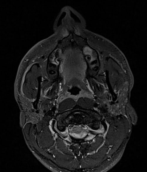
STIR axial sequence used in face imaging
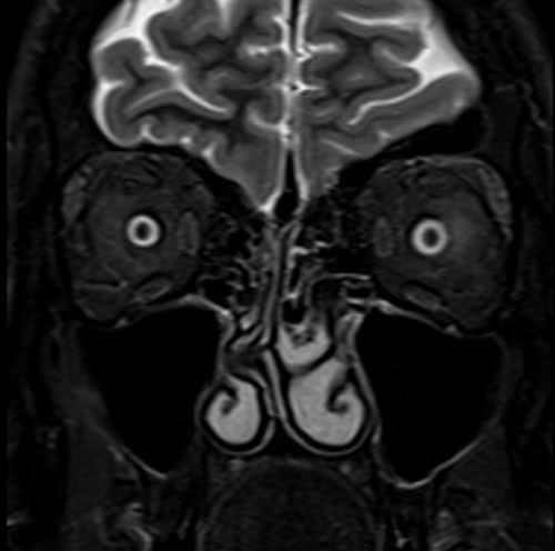
STIR coronal sequence used in orbits imaging
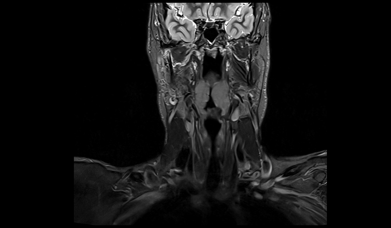
STIR coronal sequence used in neck imaging
STIR sagittal sequence used in C spine imaging

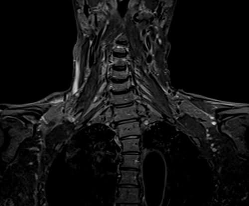
STIR coronal sequence used in b plexus imaging
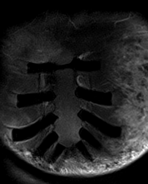
STIR coronal sequence used in sternum imaging
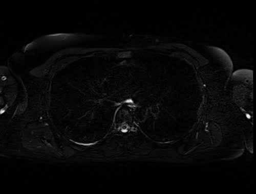
STIR axial sequence used in chest imaging
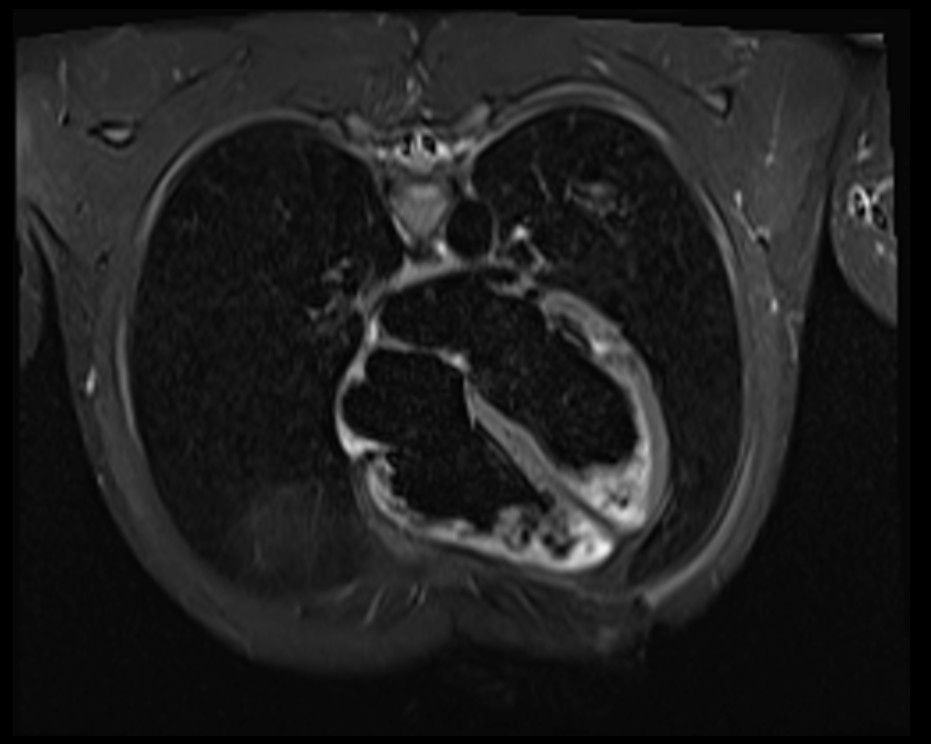
STIR 4 chamber image of heart
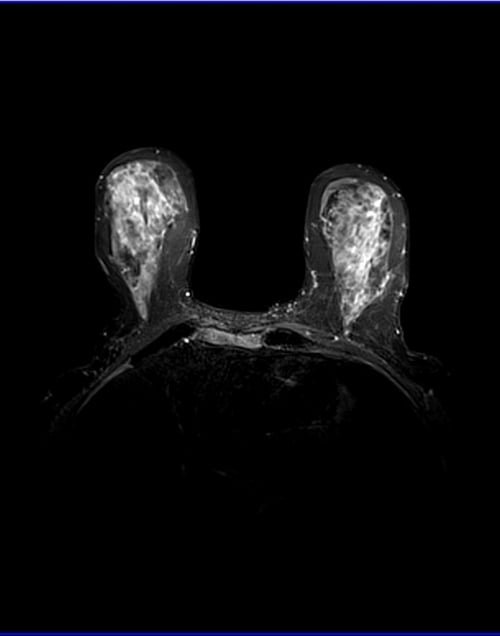
STIR axial sequence used in breast imaging
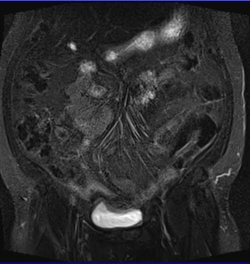
STIR coronal sequence used in abdominal imaging
STIR axial sequence used in pelvic imaging

STIR sagittal sequence used in L spine imaging

STIR coronal sequence used in SIJ imaging

STIR axial sequence used in L plexus imaging

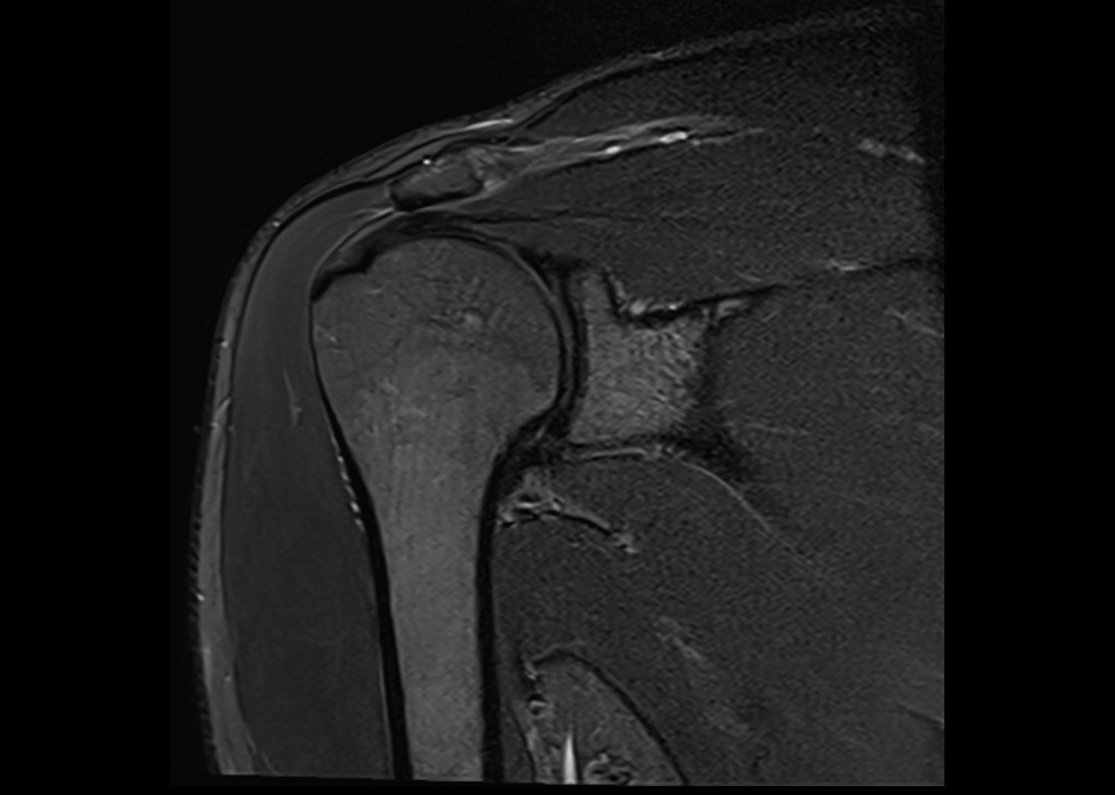
STIR coronal sequence used in shoulder imaging
STIR coronal sequence used in elbow imaging

STIR coronal sequence used in wrist imaging

STIR coronal sequence used in finger imaging

STIR coronal sequence used in hips imaging

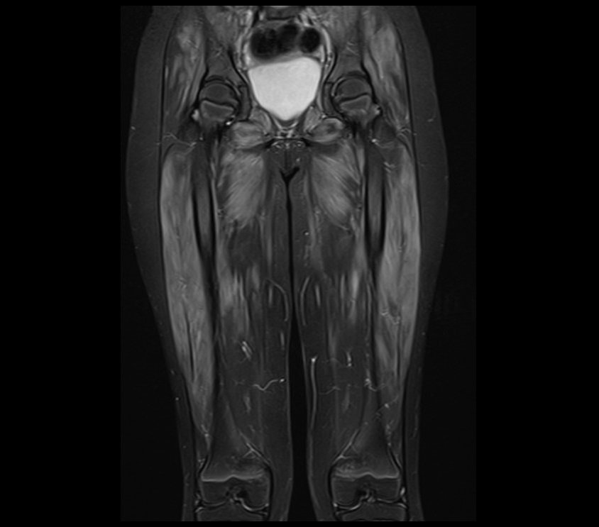
STIR coronal sequence used in thigh imaging
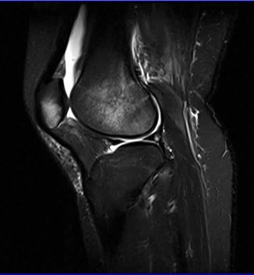
STIR sagittal sequence used in knee imaging
STIR coronal sequence used in lower legs imaging

STIR sagittal sequence used in ankle imaging

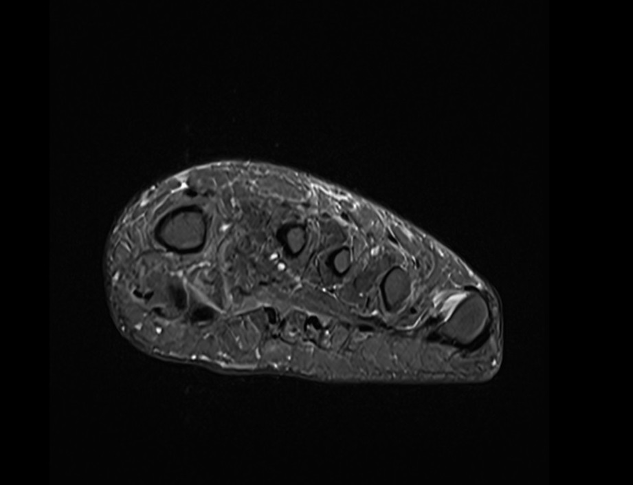
STIR axial sequence used in foot imaging
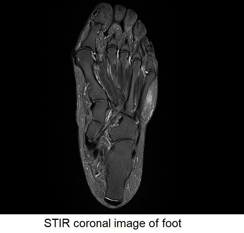
STIR coronal sequence used in foot imaging
