MRI Cerebral Aneurysm (Berry Aneurysm)
Berry aneurysms, also known as saccular aneurysms, are balloon-like bulges of blood vessels that occur in the brain. They are typically small, rounded, and form at the branching points of arteries. These aneurysms are a significant concern because they can lead to serious or life-threatening bleeding if they rupture.
Causes
The exact cause of berry aneurysms is not fully understood, but they are believed to result from a combination of genetic and environmental factors. Risk factors include:
- Genetic predisposition: Some people inherit a tendency to develop aneurysms.
- High blood pressure: Chronic hypertension can weaken blood vessel walls.
- Smoking: Tobacco use is a significant risk factor.
- Age: They are more common in adults than in children.
- Sex: Females are more likely to develop cerebral aneurysms than males.
Symptoms
Most berry aneurysms do not produce symptoms unless they leak or rupture. When a berry aneurysm ruptures, it leads to a subarachnoid hemorrhage, which can cause sudden symptoms such as:
- Sudden, severe headache (often described as “the worst headache” ever experienced)
- Nausea and vomiting
- Stiff neck
- Sensitivity to light
- Seizures
- Loss of consciousness
Unruptured aneurysms might occasionally cause symptoms if they press on brain structures or nerves, potentially leading to pain above and behind one eye, a dilated pupil, change in vision, or double vision.
Diagnosis
Berry aneurysms are typically diagnosed using imaging techniques that allow the radiologist to see the blood vessels of the brain. Common methods include:
- Computed Tomography (CT) Scan: Especially useful if the aneurysm has ruptured and there is bleeding in the brain.
- Magnetic Resonance Imaging (MRI): Provides high-resolution images of the brain and blood vessels.
Treatment
Treatment for berry aneurysms depends on the size, location, and risk of rupture. Options include:
- Surgical Clipping: This involves placing a tiny metal clip at the base of the aneurysm to isolate it from normal blood circulation, which helps prevent rupture.
- Endovascular Coiling: This minimally invasive procedure involves threading a catheter through the blood vessels to the aneurysm site, where coils are deployed to promote blood clotting and thus seal off the aneurysm.
- Observation: Small aneurysms that are not causing symptoms and have a low risk of rupture may be monitored with regular imaging studies.
MRI Appearance of Berry Aneurysms
Time-of-Flight (TOF) MRI Appearance
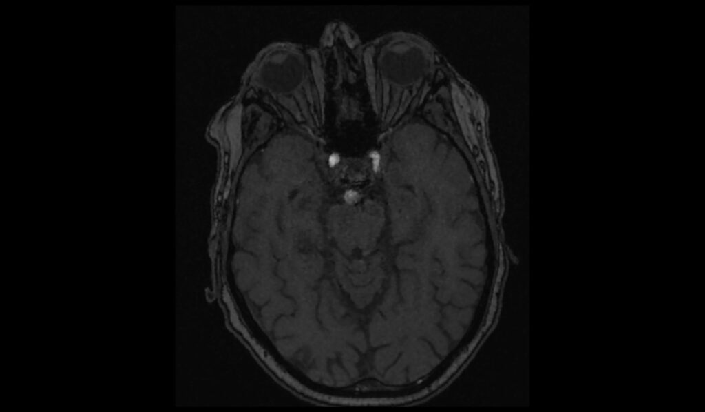
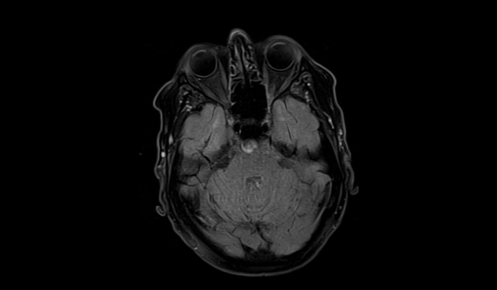
Time-of-Flight (TOF) MRI is particularly useful for visualizing the blood flow dynamics within Berry aneurysms. TOF MRI exploits the flow-related enhancement of moving blood; thus, these aneurysms typically appear as intensely bright signals due to the inflow of unsaturated spins from the circulating blood. This method can effectively delineate the aneurysm from adjacent structures and is beneficial for assessing the aneurysm neck and the relationship with parent vessels without the need for contrast agents.
T2 MRI Appearance
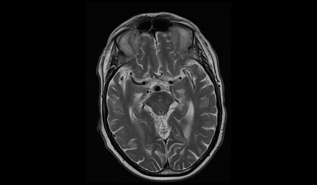
In T2-weighted MRI images, Berry aneurysms generally present as hypointense or isointense compared to cerebrospinal fluid. The characteristic flow void of circulating blood within the aneurysm appears dark on T2 imaging, while a thrombus within the aneurysm may cause variable signal intensity. Acute thrombi appear hypointense, whereas older thrombi that have undergone organization can appear hyperintense, giving a mixed signal pattern.
FLAIR MRI Appearance
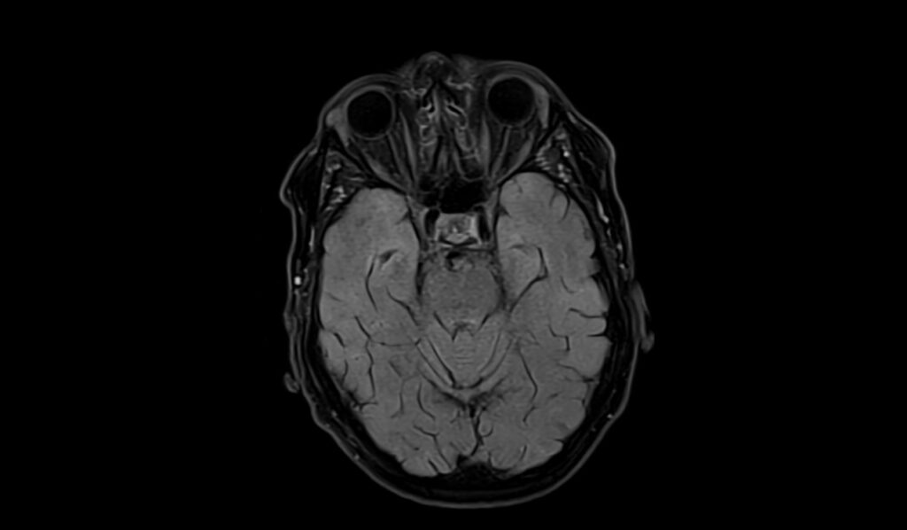
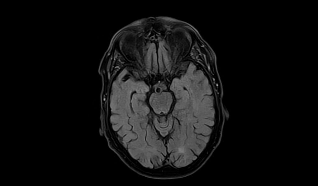
On Fluid-Attenuated Inversion Recovery (FLAIR) MRI sequences, Berry aneurysms are usually observed as signal voids, similar to their appearance on T2 images.
T1 MRI Appearance
On T1-weighted MRI images, Berry aneurysms generally appear isointense or slightly hypointense relative to the brain parenchyma. The specific appearance can vary depending on the presence of thrombosis or calcification within the aneurysm
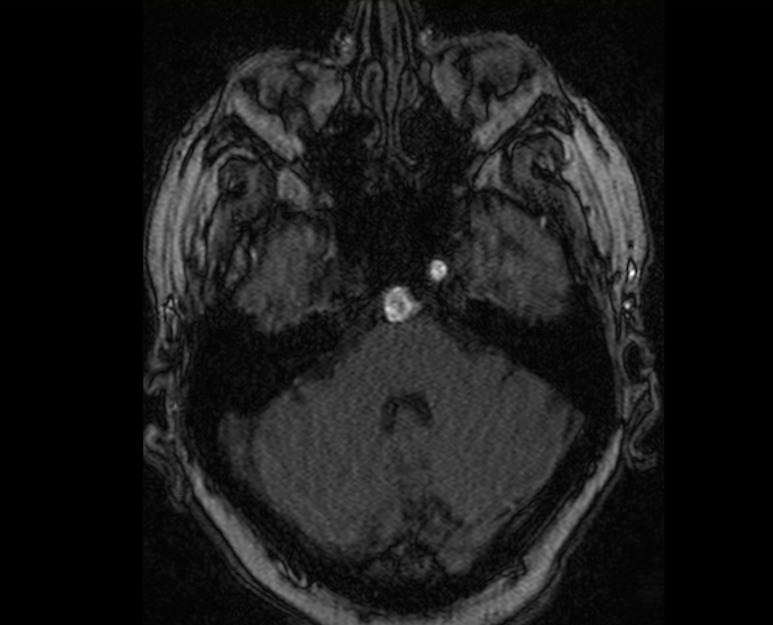
TOF axial image shows Berry Aneurysms


T2 axial image shows Berry Aneurysms


FLAIR axial image shows Berry Aneurysms
Case study 2
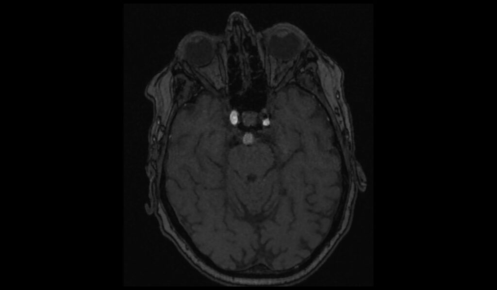
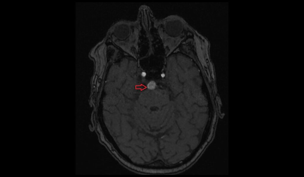
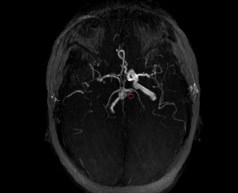
TOF MIP axial image shows cerebral Aneurysm
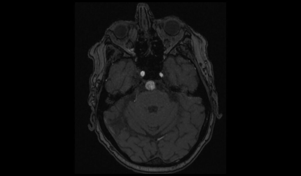
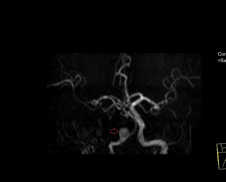
TOF MIP coronal image shows cerebral Aneurysm
TOF MIP axial image shows cerebral Aneurysm
References
- Hacein-Bey, L., & Provenzale, J. M. (2011). Current imaging assessment and treatment of intracranial aneurysms. American Journal of Roentgenology, 196(1). https://doi.org/10.2214/AJR.10.5329
- Hitchcock, E., & Gibson, W. T. (2017). A review of the genetics of intracranial berry aneurysms and implications for genetic counseling. Journal of Genetic Counseling, 26(1), 21-31. https://doi.org/10.1007/s10897-016-0029-8
- Maupu, C., Lebas, H., & Boulaftali, Y. (2022). Imaging modalities for intracranial aneurysm: More than meets the eye. Frontiers in Cardiovascular Medicine, 9, Article 793072. https://doi.org/10.3389/fcvm.2022.793072
- Phadke, R. V., Singh, V., Balaguruswamy, M. M., Udiya, A., Shetty, G. S., Prasad, S. N., Mittal, S., Chauhan, G., Dhull, V., & Neyaz, Z. (2020). Getting more out of follow-up three-dimensional time-of-flight magnetic resonance angiography in endovascularly treated intracranial aneurysms. Asian Journal of Neurosurgery, 15(4), 889–898. https://doi.org/10.4103/ajns.AJNS_374_20