Brain metastases
Brain metastases, also known as secondary brain tumors, are cancerous tumors that have originated in another part of the body and spread (metastasized) to the brain. These tumors are distinct from primary brain tumors, which originate within the brain itself. Brain metastases are a common complication of advanced cancer and can arise from various primary cancer types, such as lung, breast, melanoma, colon, and kidney cancers, among others.
Metastatic cancer cells can travel through the bloodstream or lymphatic system from their original site to the brain. Once in the brain, they can grow and form new tumors. Brain metastases are more common than primary brain tumors and are often detected in individuals with a history of cancer or in those undergoing cancer treatment.
Symptoms of brain metastases can vary and may include headaches, seizures, changes in cognitive function, weakness or numbness in specific body parts, difficulty with speech or vision, and personality changes. The symptoms depend on the location and size of the metastatic tumors.
The diagnosis of brain metastases typically involves medical imaging such as MRI or CT scans, which can reveal the presence, location, and number of tumors in the brain. Treatment options may include surgery, radiation therapy, chemotherapy, targeted therapies, and palliative care to manage symptoms and improve quality of life.
MRI appearance
The MRI appearance of brain metastases depends on factors such as the primary cancer type, size and location of metastases. The appearance of brain metastasis on various MRI sequences includes:
- T1-Weighted Images (T1WI): Brain metastases often appear hypointense on T1-weighted images. The signal intensity of metastatic lesions is generally lower than that of surrounding normal brain tissue. This is due to the increased cellularity and compactness of cancerous tissue in comparison to the brain parenchyma.
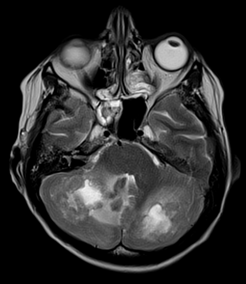
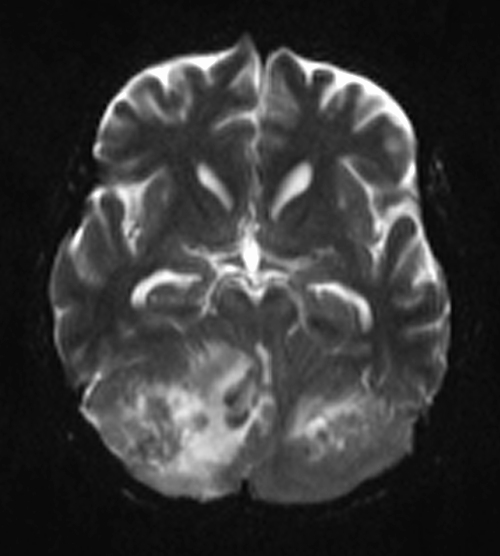
- T2-Weighted Images (T2WI): Brain metastases can vary in appearance on T2-weighted images. They often appear hyperintense due to their relatively high water content. The hyperintensity is caused by the increased extracellular space and edema associated with the tumor.
- Fluid-Attenuated Inversion Recovery (FLAIR): Metastases typically appear hyperintense on FLAIR images due to the edema and increased water content surrounding the tumor. FLAIR sequences effectively suppress the cerebrospinal fluid signal, enhancing the visibility of peritumoral edema.
- Diffusion-Weighted Imaging (DWI): Metastatic lesions can show variable signal intensity on DWI depending on factors such as cellularity and restricted diffusion. They may appear hyperintense due to reduced diffusion caused by increased cellularity and cellular membrane structures.
- Post-Contrast T1-Weighted Images: After the administration of contrast material (gadolinium-based), metastatic lesions typically enhance. The degree of enhancement can vary, and lesions may appear as hyperintense areas due to the increased permeability of blood vessels within the tumor. The enhancement pattern can provide insights into the tumor’s vascularity
T2 Axial

T1 post contrast Axial
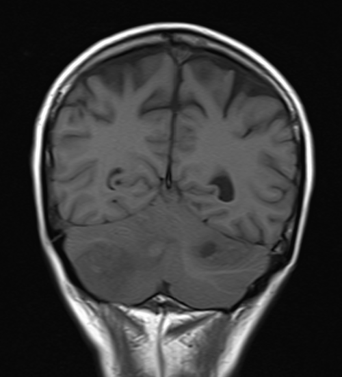
T1 Coronal Pre Contrast
AXIal DWI B0

AXIal DWI B1000

AXIal ADC MAP

T1 post contrast coronal
References
Patchell, R. A., Tibbs, P. A., Walsh, J. W., Dempsey, R. J., Maruyama, Y., Kryscio, R. J., … & Young, B. (1990). A randomized trial of surgery in the treatment of single metastases to the brain. New England Journal of Medicine, 322(8), 494-500.
Nayak, L., Lee, E. Q., Wen, P. Y. (2012). Epidemiology of brain metastases. Current Oncology Reports, 14(1), 48-54.
Barnholtz-Sloan, J. S., Sloan, A. E., Davis, F. G., Vigneau, F. D., Lai, P., Sawaya, R. E. (2004). Incidence proportions of brain metastases in patients diagnosed (1973 to 2001) in the Metropolitan Detroit Cancer Surveillance System. Journal of Clinical Oncology, 22(14), 2865-2872.
Meyer, M. A., & King, A. D. (2014). The staging of cancer. Cancer Imaging: Lung and Breast Carcinomas, 1-14.
Lin, N. U., Lee, E. Q., Aoyama, H., Barani, I. J., Baumert, B. G., Brown, P. D., … & Aoyama, H. (2019). Response assessment criteria for brain metastases: proposal from the RANO group. The Lancet Oncology, 16(6), e270-e278.