Arachnoid cyst MRI
An arachnoid cyst is a cerebrospinal fluid (CSF)-filled sac located between the brain or spinal cord and the arachnoid membrane, one of the three meninges that cover the brain and spinal cord. These cysts are typically benign and non-cancerous but can cause symptoms if they grow large enough to compress surrounding neural structures.
Causes
The exact cause of arachnoid cysts is often unknown, but they are generally categorized into two types based on their origin:
- Primary (Congenital): These cysts are present at birth and arise due to developmental abnormalities during the formation of the brain and spinal cord.
- Secondary (Acquired): These cysts develop later in life due to factors such as head injury, meningitis, tumors, or brain surgery.
Symptoms
Many arachnoid cysts are asymptomatic and discovered incidentally during imaging studies for other conditions. However, symptomatic cysts can lead to various neurological symptoms, which depend on their size and location. Common symptoms include:
- Headache
- Nausea and vomiting
- Seizures
- Hydrocephalus (accumulation of CSF in the brain)
- Developmental delays in children
- Balance and coordination problems
- Hearing or vision disturbances
- Cognitive and behavioral changes
Diagnosis
The diagnosis of arachnoid cysts typically involves imaging studies to visualize the cyst and its impact on surrounding brain structures. Common diagnostic methods include:
- Magnetic Resonance Imaging (MRI): Provides detailed images of the brain and spinal cord, helping to identify the cyst’s size, location, and effect on adjacent tissues.
- Computed Tomography (CT) Scan: Useful for identifying cysts, especially in emergency settings. It provides less detail than MRI but is quicker.
- Ultrasound: Occasionally used in infants with open fontanelles (soft spots) to visualize cysts.
Treatment
The treatment approach for arachnoid cysts depends on the presence and severity of symptoms. Options include:
- Observation: Asymptomatic or minimally symptomatic cysts are often monitored with regular imaging to ensure they do not grow or cause problems.
- Medications: Symptomatic treatment for headaches, seizures, or other specific symptoms.
- Surgical Intervention: Considered for cysts causing significant symptoms or complications. Surgical options include:
- Endoscopic Fenestration: A minimally invasive procedure where small openings are created in the cyst to allow CSF to flow into the normal pathways.
- Microsurgical Removal: Complete or partial removal of the cyst, usually for more accessible or problematic cysts.
- Shunting: Involves inserting a shunt to divert CSF from the cyst to another body cavity (e.g., the abdomen) to relieve pressure.
MRI appearance of Arachnoid cyst
T1-Weighted Imaging
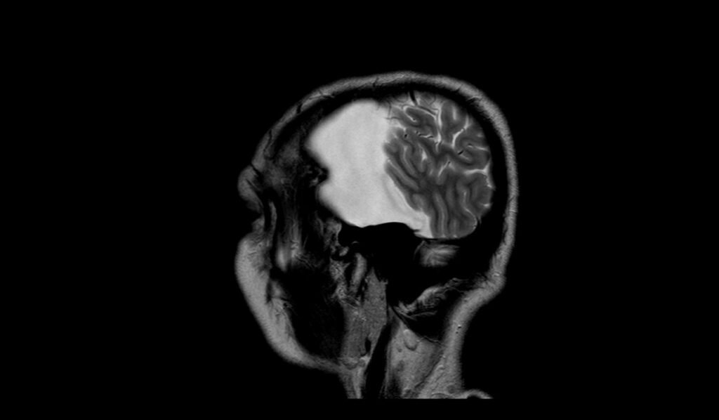
On T1-weighted MRI, arachnoid cysts typically appear as well-defined, hypointense (dark) areas relative to brain tissue. The cysts are filled with cerebrospinal fluid (CSF), which has a low signal on T1 images, making the cysts distinguishable from the surrounding brain structures.
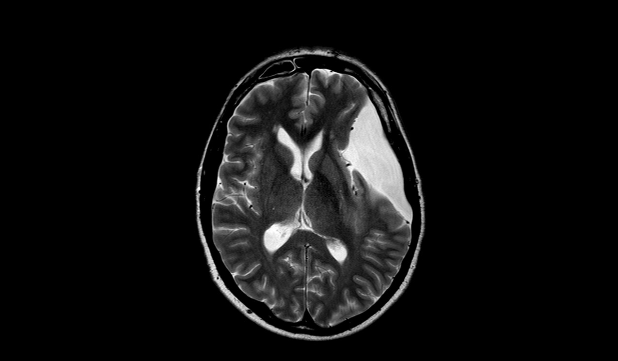
T2-Weighted Imaging
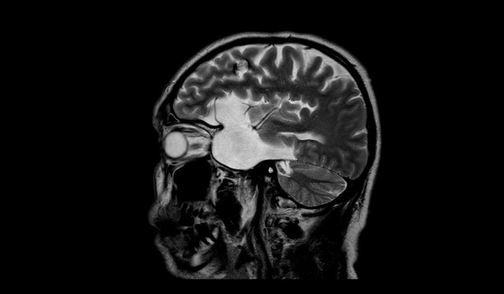
On T2-weighted MRI, arachnoid cysts appear hyperintense (bright) due to the high water content of the CSF within the cyst. This bright signal helps to clearly delineate the cyst from the adjacent brain parenchyma, which has a relatively lower signal intensity.
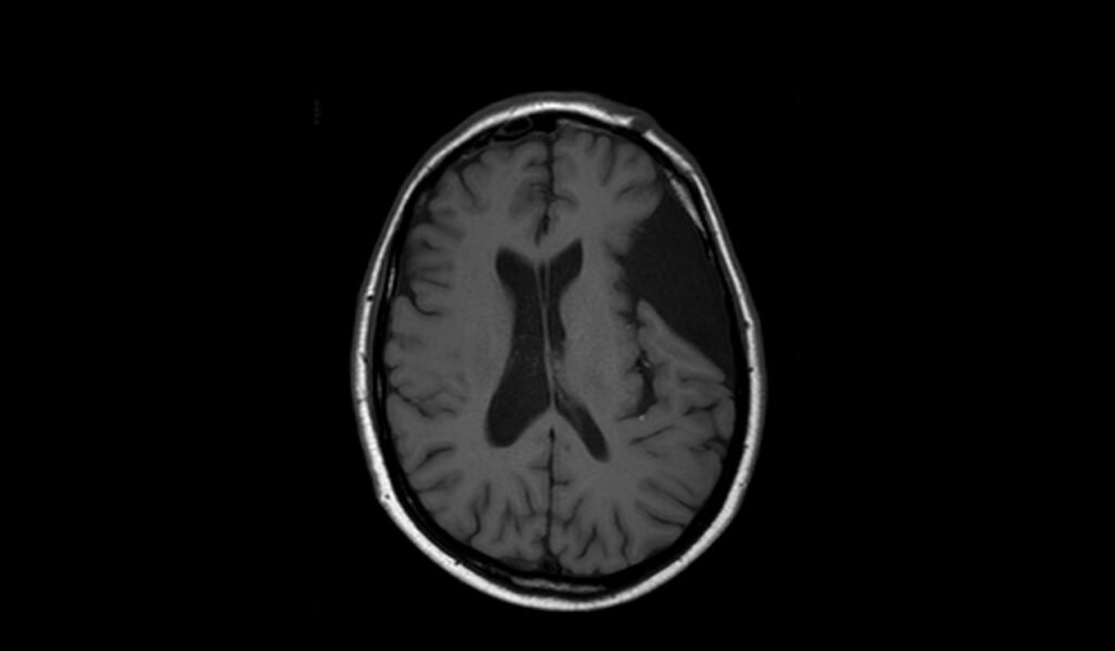
FLAIR (Fluid-Attenuated Inversion Recovery)
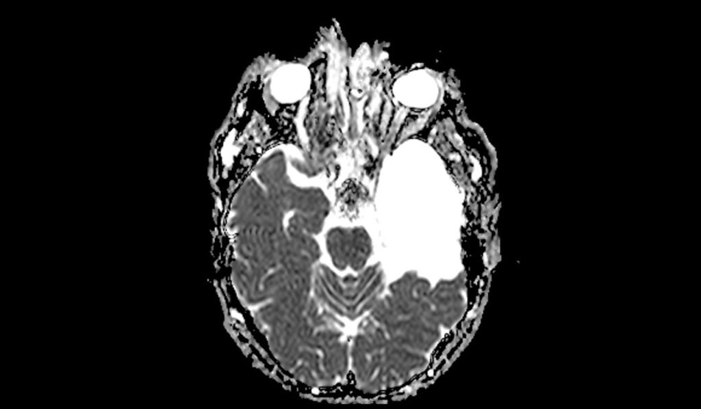
FLAIR images show arachnoid cysts as hypointense (dark) areas, similar to the signal of CSF. Unlike other fluid-filled lesions, arachnoid cysts do not show a bright signal on FLAIR, which helps in differentiating them from other pathologies such as tumors or abscesses that may have a different signal intensity.
DWI (Diffusion-Weighted Imaging) and ADC (Apparent Diffusion Coefficient)
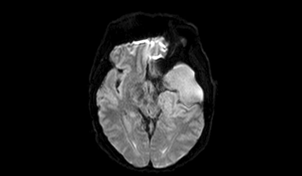
On DWI (with b0 and b1000) and ADC maps, arachnoid cysts typically exhibit no diffusion restriction. They appear hypointense on DWI and hyperintense on ADC, similar to normal CSF. This lack of diffusion restriction helps differentiate arachnoid cysts from other lesions that may restrict diffusion, such as abscesses or some tumors.
T2 axial image shows Arachnoid cyst


FLAIR axial image shows Arachnoid cyst
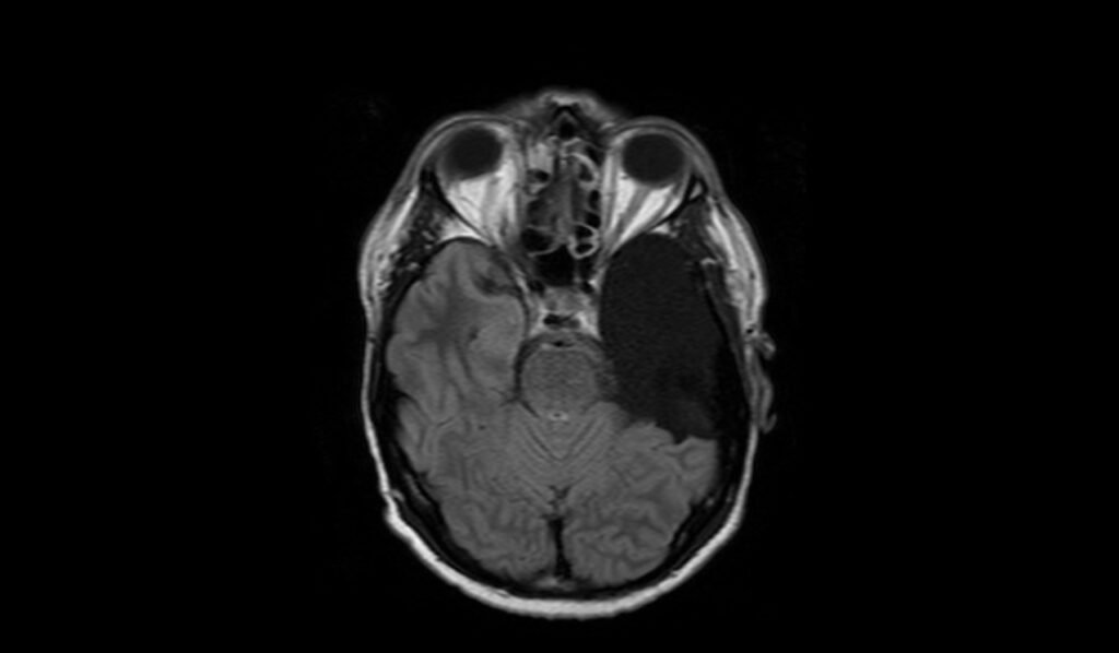


T1 axial images shows Arachnoid cyst

T2 sagittal images shows Arachnoid cyst

DWI b0 images shows Arachnoid cyst
DWI b1000 images shows Arachnoid cyst

DWI b1000 images shows Arachnoid cyst
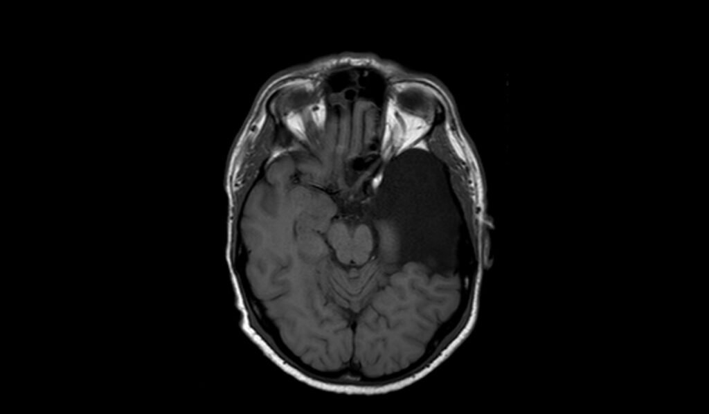
T1 post contrast axial images shows Arachnoid cyst
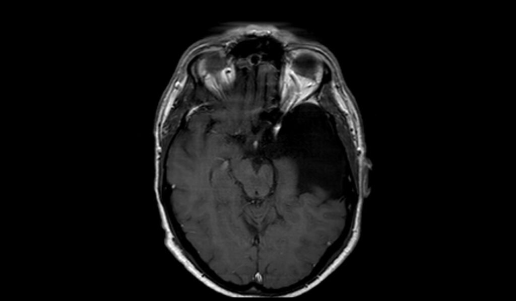
T1 post contrast coronal images shows Arachnoid cyst

References
- White, M. L., & Das, J. M. (2024, February 2). Arachnoid Cysts. In StatPearls [Internet]. StatPearls Publishing. Retrieved from https://www.ncbi.nlm.nih.gov/books/NBK532297/
- Mavroidis, P., Roka, V., Kostopoulos, S., Batsikas, G., & Lavdas, E. (2016). Arachnoid cysts: The role of the BLADE technique. Hippokratia, 20(3), 244–248. https://doi.org/10.1186/PMC5654446
- Dlaka, D., Raguž, M., Muller, D., Romić, D., Almahariq, F., Dlaka, J., Kaštelančić, A., & Chudy, D. (2019). Intraparenchymal supratentorial arachnoid cyst: a case report. Egyptian Journal of Neurosurgery, 34, Article 28. https://doi.org/10.1186/s41984-019-0034-0