MRI Anterior cruciate ligament (ACL) tear
The anterior cruciate ligament (ACL) is one of the key ligaments that help stabilize the knee joint. An ACL tear is a common knee injury, especially among athletes. It occurs when the ACL is stretched beyond its limits or torn.
Causes
ACL tears often occur due to:
- Sudden Stops and Changes in Direction: Rapid changes in movement, common in sports like soccer, basketball, and football.
- Landing Incorrectly from a Jump: Incorrect landing mechanics after a jump.
- Direct Blow to the Knee: Direct contact or collision, often in contact sports.
- Overextension: Hyperextending the knee.
Symptoms
Common symptoms of an ACL tear include:
- A Loud “Pop” Sound: Often reported at the time of injury.
- Severe Pain: Immediate and intense pain in the knee.
- Swelling: Swelling begins within a few hours of the injury.
- Instability: Feeling of the knee “giving way” when putting weight on it.
- Limited Range of Motion: Difficulty in fully bending or straightening the knee.
- Tenderness: Tenderness along the joint line
Diagnosis
Diagnosing an ACL tear involves a combination of:
- Physical Examination: A doctor will assess knee stability and pain levels through specific maneuvers, such as the Lachman test or the pivot-shift test.
- Imaging Tests:
- Magnetic Resonance Imaging (MRI): The most effective imaging technique for confirming an ACL tear, showing the extent of the injury and any associated damage to other knee structures.
- X-rays: Usually done to rule out bone fractures.
Treatment
Treatment options for an ACL tear depend on the severity of the injury, the patient’s activity level, and overall health. They include:
Non-Surgical Treatment:
- Physical Therapy: To restore knee function and strengthen surrounding muscles.
- Bracing: Wearing a knee brace to provide stability.
Surgical Treatment:
- ACL Reconstruction Surgery: Involves replacing the torn ligament with a graft, usually from the patient’s own hamstring or patellar tendon, or from a donor. Surgery is often recommended for active individuals or athletes to regain full knee function.
MRI Appearance of Patellar Dislocation
T1 weighted Sequence
- Dislocation: The patella appears out of its normal position, often laterally displaced.
- Bone Contusions: There may be visible bone marrow edema in the lateral femoral condyle and the medial patella as areas of low signal intensity.
- Medial Retinaculum and Medial Patellofemoral Ligament (MPFL): Potential tears or injuries might appear as irregularities or discontinuities in these structures.
Proton Density (PD) Fat Saturation (Fat Sat) Sequence
- Dislocation: The patella’s abnormal position will be clearly seen, usually with lateral displacement.
- Bone Marrow Edema: Enhanced visualization of bone marrow edema in the lateral femoral condyle and medial patella as high signal intensity areas due to the fat suppression technique.
- Soft Tissue Structures: Injuries to the MPFL and medial retinaculum appear more conspicuous, showing as high signal intensity due to the increased fluid content from acute injury.
- Joint Effusion: Increased fluid within the joint space will be seen as high signal intensity.
Short Tau Inversion Recovery (STIR) Sequence
- Dislocation: The patella remains visibly out of place.
- Bone Marrow Edema: Enhanced detection of bone marrow edema in the affected bones due to the high sensitivity of STIR to fluid, showing as very high signal intensity areas.
- Soft Tissue Edema: The presence of edema in the surrounding soft tissues, including the medial retinaculum and MPFL, will appear as areas of high signal intensity.
- Joint Effusion: Like in PD Fat Sat, joint effusion will be prominently seen as high signal intensity.
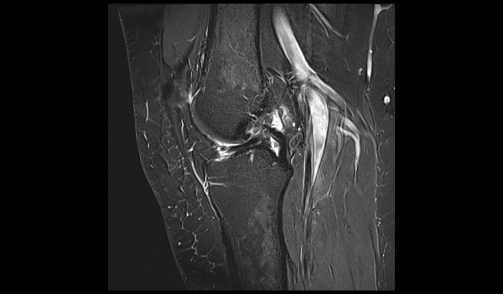
STIR sagittal image shows Anterior Cruciate Ligament (ACL) Tear

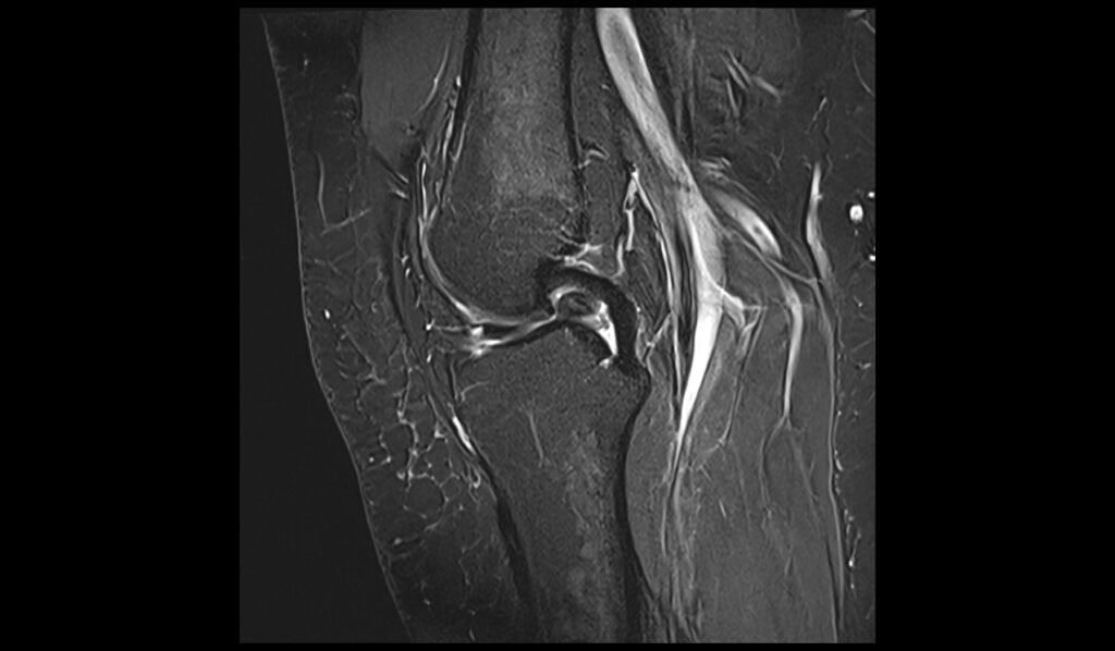
T1 TSE sagittal image shows Anterior Cruciate Ligament (ACL) Tear


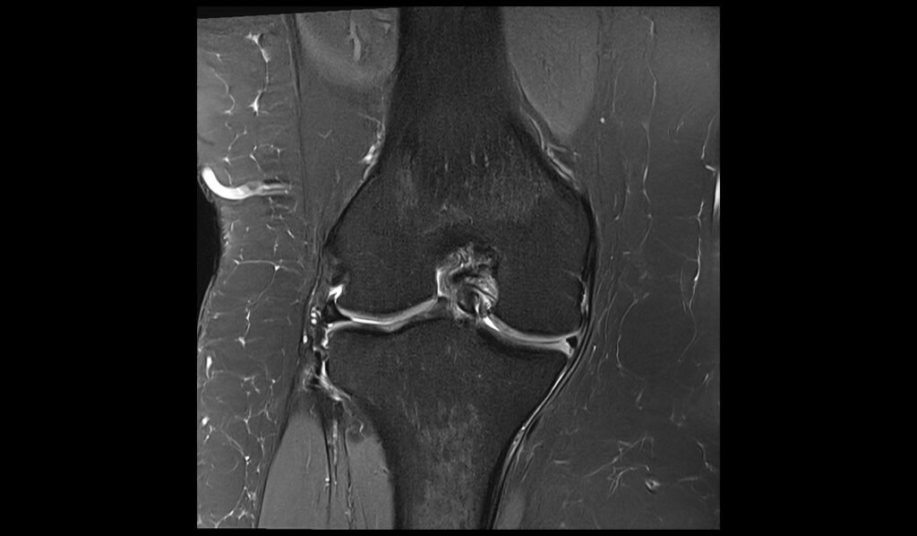
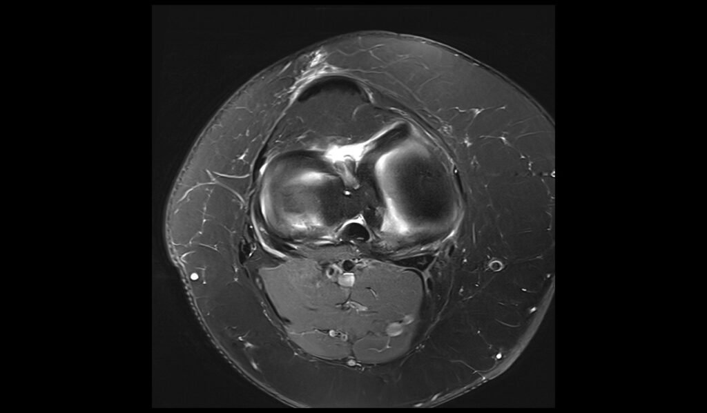
PD fat saturated coronal shows Anterior Cruciate Ligament (ACL) Tear
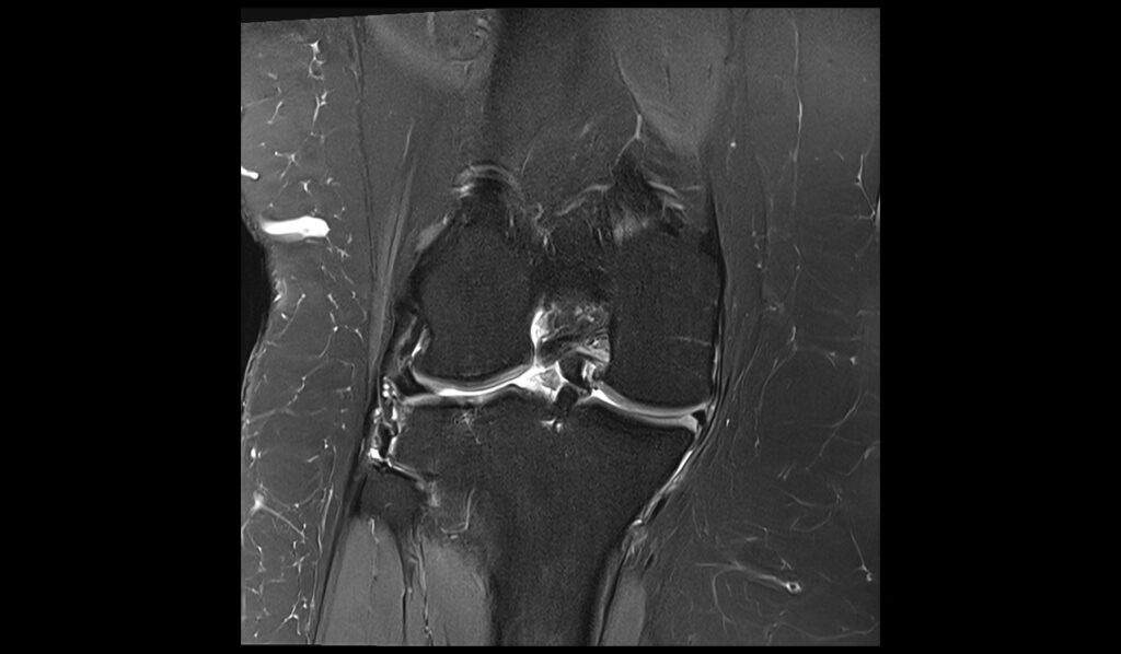

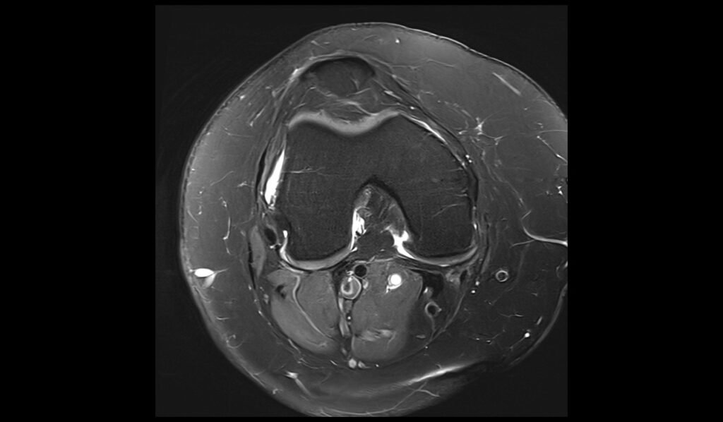
PD fat saturated axial image showsAnterior Cruciate Ligament (ACL) Tear


References
- Ng, W. H. A., Griffith, J. F., Hung, E. H. Y., Paunipagar, B., Law, B. K. Y., & Yung, P. S. H. (2011). Imaging of the anterior cruciate ligament. World Journal of Orthopedics, 2(8), 75-84. https://doi.org/10.5312/wjo.v2.i8.75
- Prince, J. S., Laor, T., & Bean, J. A. (2005). MRI of anterior cruciate ligament injuries and associated findings in the pediatric knee: Changes with skeletal maturation. American Journal of Roentgenology, 184(4), 1064-1069. https://doi.org/10.2214/ajr.184.4.01841064
- Guenoun, D., Le Corroller, T., Amous, Z., Pauly, V., Sbihi, A., & Champsaur, P. (2012). The contribution of MRI to the diagnosis of traumatic tears of the anterior cruciate ligament. Diagnostic and Interventional Imaging, 93(5), 331-341. https://doi.org/10.1016/j.diii.2012.02.003