Acute Pontine Infarct MRI
An acute pontine infarct is a type of ischemic stroke that occurs specifically in the pons, which is part of the brainstem. The brainstem is crucial for several basic bodily functions, including breathing, heart rate regulation, and consciousness.
Causes
Acute pontine infarcts are typically caused by the interruption of blood flow to the pons. The main causes include:
- Atherosclerosis: The buildup of plaques in arteries, which can reduce or block blood flow.
- Small Vessel Disease: This involves the narrowing of small blood vessels within the brain, which is common in conditions like hypertension and diabetes.
- Embolism: Blood clots from other parts of the body (like the heart in atrial fibrillation) can travel to the brain and block a blood vessel.
- Arterial Dissection: A tear in the lining of an artery, which can block blood flow or reduce it significantly.
Symptoms
The symptoms of an acute pontine infarct can be severe and sudden, reflecting the critical functions managed by the pons. They include:
- Weakness or paralysis: Typically on one side of the body.
- Difficulty speaking or swallowing.
- Double vision or other vision problems.
- Vertigo and imbalance.
- Altered consciousness, ranging from slight confusion to coma.
Diagnosis
Diagnosis of an acute pontine infarct involves several steps:
- Medical History and Physical Examination: To assess symptoms and check for risk factors.
- Imaging Tests: Magnetic resonance imaging (MRI) is the most sensitive test for diagnosing brainstem strokes, as it can detect changes in brain tissue and blood flow.
- Computed Tomography (CT) Scan: Useful for quickly ruling out a hemorrhagic stroke.
Treatment
The treatment for an acute pontine infarct aims to restore blood flow and minimize brain damage:
- Thrombolytic Therapy: Administering drugs like tissue plasminogen activator (tPA) to dissolve clots. This treatment is time-sensitive, usually within a few hours of symptom onset.
- Anticoagulant and Antiplatelet Therapy: Medications such as warfarin, aspirin, or clopidogrel to prevent new clots.
- Control
MRI appearance of Acute Pontine Infarct
MRI T2 Appearance of Acute Pontine Infarct
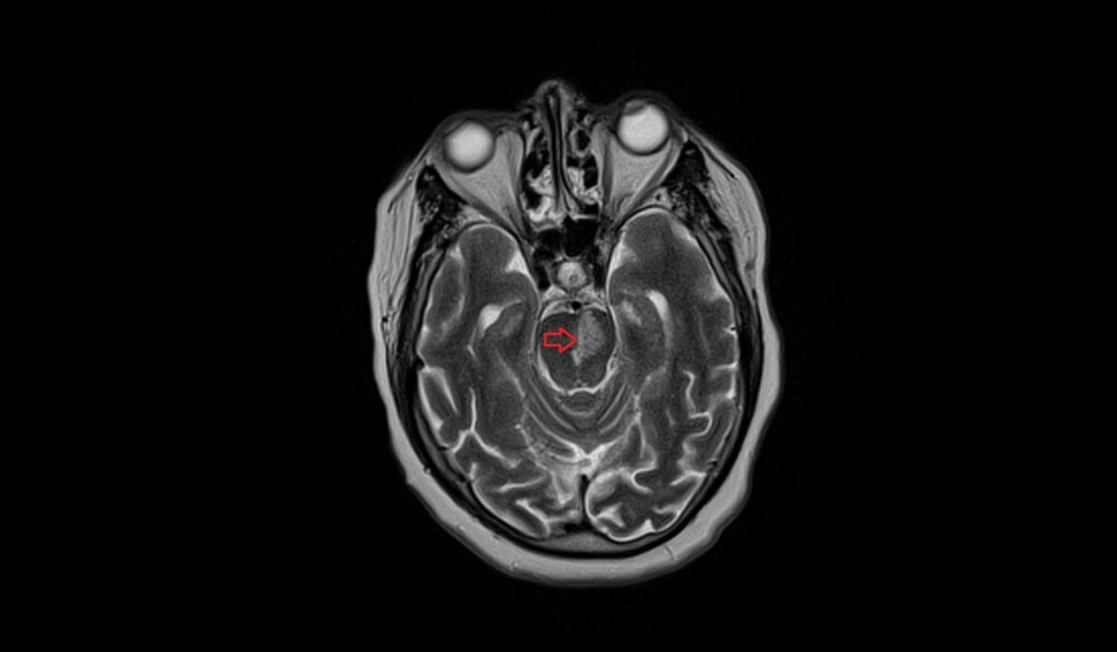
T2-weighted MRI images of an acute pontine infarct usually reveal a hyperintense (bright) signal in the infarcted region. This hyperintensity corresponds to the increased water content and edema associated with the acute phase of the infarct. T2-weighted imaging is particularly sensitive to these changes, making it effective for identifying the affected area. The bright signal on T2 images helps to differentiate the infarcted tissue from the surrounding normal brain parenchyma.
MRI FLAIR Appearance of Acute Pontine Infarct
FLAIR (Fluid-Attenuated Inversion Recovery) sequences enhance the visibility of lesions by suppressing the CSF signal, making them useful for detecting infarcts. In an acute pontine infarct, FLAIR images typically show a hyperintense (bright) signal in the pons, similar to T2-weighted images. This hyperintensity highlights the infarcted area, allowing for better visualization against the background of suppressed CSF. FLAIR is particularly valuable for identifying small infarcts and assessing the extent of ischemic damage.
MRI DWI b0, b1000, and ADC Appearance of Acute Pontine Infarct
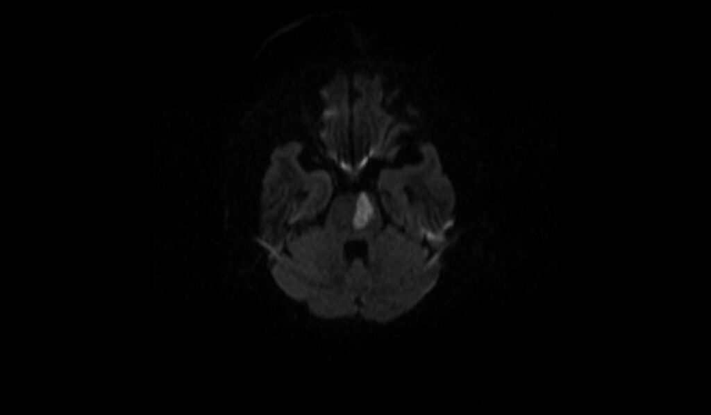
Diffusion-weighted imaging (DWI) is highly sensitive for detecting acute ischemic strokes. On DWI with b1000, an acute pontine infarct appears as a hyperintense (bright) area due to restricted diffusion of water molecules within the ischemic tissue. The b0 image serves as a reference with normal signal intensity. The apparent diffusion coefficient (ADC) map typically shows a corresponding hypointense (dark) signal in the infarcted area, reflecting the restricted diffusion. This combination of DWI hyperintensity and ADC hypointensity is characteristic of acute infarction, providing crucial information for early diagnosis and treatment planning.
MRI T1 Appearance of Acute Pontine Infarct
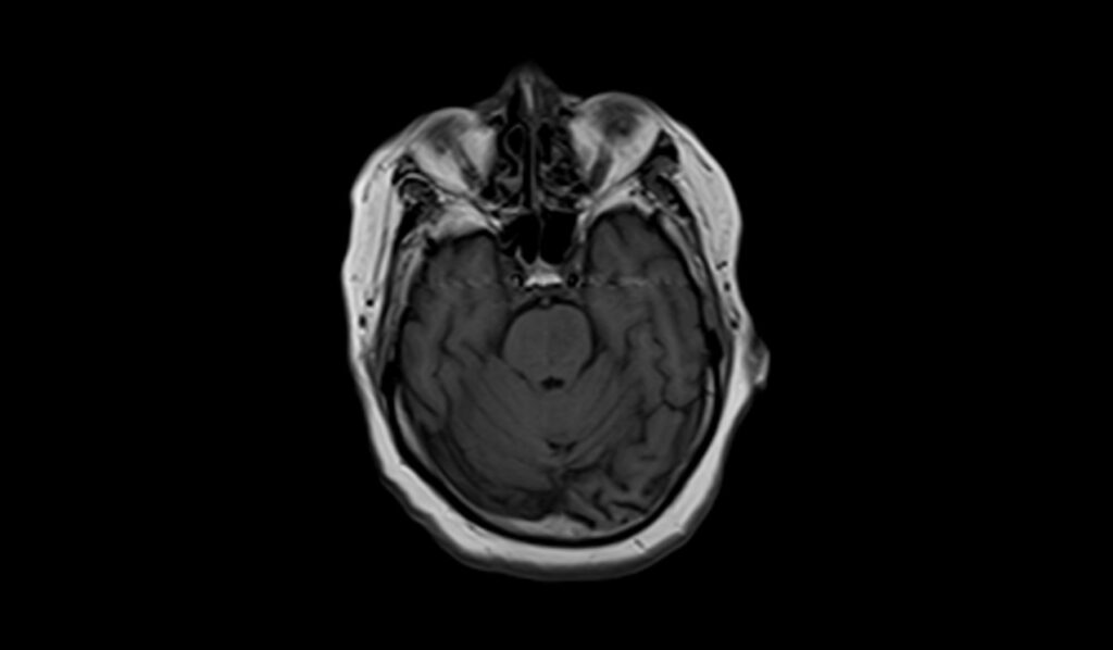
In the case of an acute pontine infarct, T1-weighted MRI images typically show a hypointense (dark) signal in the affected area of the pons. This hypointensity is due to the early presence of ischemic tissue, which lacks the normal signal intensity of healthy brain tissue. T1-weighted imaging is useful for assessing the anatomical detail and delineating the extent of the infarct within the pons, although it may not be as sensitive as other sequences for detecting acute changes.
T2 axial images shows Acute Pontine Infarct (stroke)
FLAIR axial image shows Acute Pontine Infarct (stroke)

T1 axial image shows shows Acute Pontine Infarct (stroke)
DWI b0 image shows Acute Pontine Infarct (stroke)

DWI b500 image shows Acute Pontine Infarct (stroke)
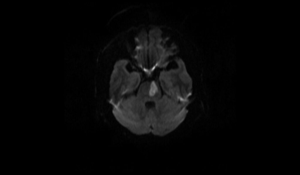
DWI b1000 image shows Acute Pontine Infarct (stroke)
DWI ADC map image shows Acute Pontine Infarct (stroke)

References
- Kumral, E., Bayülkem, G., & Evyapan, D. (2002). Clinical spectrum of pontine infarction. Clinical-MRI correlations. Journal of Neurology, 249(12), 1659-1670.
- Mäntylä, R., Pohjasvaara, T., Vataja, R., Salonen, O., Aronen, H. J., Standertskjöld-Nordenstam, C.-G., Kaste, M., & Erkinjuntti, T. (2000). MRI pontine hyperintensity after supratentorial ischemic stroke relates to poor clinical outcome. Stroke, 31(3), 695-700. https://doi.org/10.1161/01.STR.31.3.695
- Vlašković, T., Georgievski Brkić, B., Stević, Z., Vukićević, M., Đurović, O., Kostić, D., Stanisavljević, N., Marinković, I., Kapor, S., & Marinković, S. (2022). Anatomic and MRI bases for pontine infarctions with patients presentation. Journal of Stroke and Cerebrovascular Diseases, 31(8), 106613. Elsevier.
- Mäntylä R, Pohjasvaara T, Vataja R, et al. MRI pontine hyperintensity after supratentorial ischemic stroke relates to poor clinical outcome. Stroke. 2000;31(3):695-700. doi:10.1161/01.STR.31.3.695
- Wu, L., Li, Y., Ye, Z., Liu, D., Dai, Z., Zhu, J., Chen, H., Li, C., Lie, C., & Jiang, Y. (2022). Site and mechanism of recurrent pontine infarction: A hospital-based follow-up study. Brain Sciences, 12(5), 520.