Acoustic neuroma MRI
Acoustic Neuroma (Vestibular Schwannoma) is a non-cancerous (benign) tumor that develops on the vestibulocochlear nerve, which connects the inner ear to the brain. This nerve is responsible for transmitting sound and balance information. Acoustic neuromas are slow-growing tumors that typically arise from Schwann cells, which produce the myelin sheath covering the nerve.
Causes of Acoustic Neuroma
The exact cause of most acoustic neuromas is unknown, but some cases are linked to a genetic disorder called neurofibromatosis type II (NF2). People with NF2 tend to develop benign tumors on multiple nerves, including both sides of the vestibulocochlear nerve.
Most acoustic neuromas occur sporadically, meaning they are not inherited or linked to a specific cause.
Symptoms of Acoustic Neuroma
Symptoms of acoustic neuroma can vary depending on the size and location of the tumor, but they typically develop slowly as the tumor grows. Common symptoms include:
- Hearing Loss: Gradual hearing loss in one ear is the most common symptom. This may be accompanied by a sensation of fullness or ringing in the ear (tinnitus).
- Tinnitus: Persistent ringing, buzzing, or hissing sound in the affected ear.
- Balance Problems: As the tumor affects the vestibular part of the nerve, dizziness, unsteadiness, and a sense of vertigo may occur.
- Facial Numbness or Weakness: In larger tumors, nearby facial nerves can be affected, leading to numbness, tingling, or muscle weakness in the face.
- Headaches: Pressure on nearby tissues or nerves may cause headaches.
- Ear Fullness: A feeling of fullness in the affected ear, without any infection.
Diagnosis of Acoustic Neuroma
Diagnosing acoustic neuroma typically involves a combination of clinical examination and imaging studies:
Hearing Test (Audiometry): A detailed hearing test is often the first step in evaluating hearing loss and can help identify the specific type of hearing loss associated with acoustic neuroma.
MRI Scan: Magnetic Resonance Imaging (MRI) with contrast is the gold standard for diagnosing acoustic neuroma. It allows for detailed imaging of the brain and inner ear structures, helping to detect even small tumors.
CT Scan: Although less sensitive than MRI, a computed tomography (CT) scan may be used if MRI is not an option.
Electronystagmography (ENG): This test may be used to evaluate balance and detect vestibular dysfunction, although it is not always necessary.
Brainstem Auditory Evoked Response (BAER): This test evaluates the hearing pathways in the brainstem and may show abnormalities when the tumor affects the auditory nerve.
Treatment of Acoustic Neuroma
Treatment depends on the size and growth of the tumor, symptoms, and overall health of the patient. Options include:
Observation (Watchful Waiting): Small, slow-growing tumors that are not causing significant symptoms may be monitored with regular MRI scans to track growth. This is often the approach for elderly patients or those for whom surgery carries high risks.
Surgery: There are several types of surgical approaches to remove the tumor, depending on its size and location. Surgery aims to remove as much of the tumor as possible while preserving hearing and facial nerve function. However, complete removal may sometimes damage nearby structures, leading to complications like hearing loss or facial nerve weakness.
- Translabyrinthine approach: Used for larger tumors or when hearing preservation is not possible.
- Retrosigmoid/suboccipital approach: Allows for the possibility of hearing preservation.
- Middle fossa approach: Used for smaller tumors, with the aim of preserving hearing.
Radiation Therapy: Stereotactic radiosurgery (such as Gamma Knife or CyberKnife) can deliver targeted radiation to shrink or stop the growth of the tumor. This option is often used for smaller tumors or for patients who are not candidates for surgery.
MRI Appearance of Acoustic Neuroma
MRI T1 Appearance of Acoustic Neuroma
On a T1-weighted MRI without contrast, an acoustic neuroma typically appears as an isointense or slightly hypointense mass relative to the surrounding brain tissue, particularly the cerebellopontine angle (CPA) or internal auditory canal (IAC). The tumor is usually not very conspicuous in this sequence due to its subtle intensity differences compared to the adjacent structures, making it difficult to differentiate from surrounding tissues without contrast enhancement.
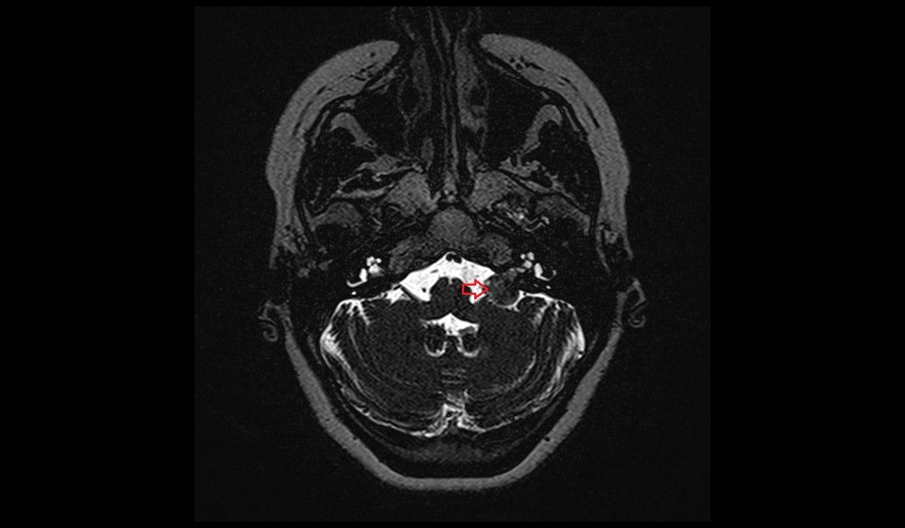
MRI SPACE Sequence Appearance of Acoustic Neuroma
In a T2-weighted SPACE (Sampling Perfection with Application-optimized Contrasts using different flip-angle Evolutions) sequence, acoustic neuromas typically appear isointense to hyperintense compared to the surrounding brain tissues. Due to the contrast between the cerebrospinal fluid (CSF), which appears bright, and the lesion, which is isointense to hyperintense, the tumor stands out more clearly in the internal auditory canal or cerebellopontine angle. The SPACE sequence provides high-resolution imaging of small structures and nerves, making it easier to distinguish even small tumors from the adjacent CSF, thereby enhancing lesion visibility.
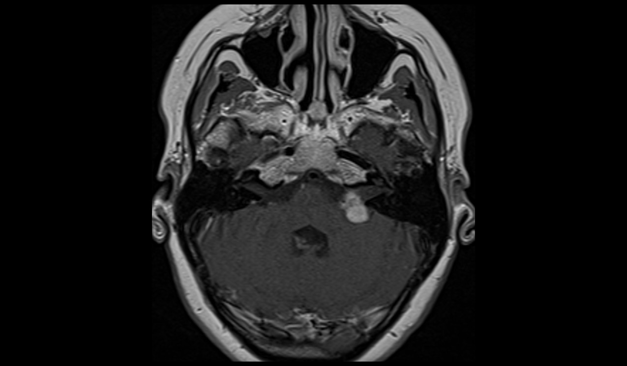
MRI T1 Post-Contrast Appearance of Acoustic Neuroma
On a T1-weighted MRI with contrast (gadolinium), acoustic neuromas show intense, homogeneous enhancement due to the vascular nature of the tumor. The contrast highlights the tumor clearly, differentiating it from surrounding tissues and allowing for more precise localization. In larger tumors, areas of necrosis or cystic degeneration may appear as non-enhancing regions, but the solid portions of the tumor enhance robustly. This post-contrast sequence is crucial for definitive diagnosis and assessing tumor size and extent.
MRI T2 SPACE axial image shows acoustic neuroma


MRI T1 TSE axial image shows acoustic neuroma


MRI T1 TSE axial post contrast image shows acoustic neuroma


MRI T1 TSE coronal post contrast image shows acoustic neuroma


References
- Greene, J., & Al-Dhahir, M. A. (2023). Acoustic Neuroma. StatPearls [Internet]. Last updated August 17, 2023. National Center for Biotechnology Information (NCBI), U.S. National Library of Medicine (NLM). Available from: https://www.ncbi.nlm.nih.gov/books/NBK441920/
- Kabashi, S., Ugurel, M. S., Dedushi, K., & Mucaj, S. (2020). The Role of Magnetic Resonance Imaging (MRI) in Diagnostics of Acoustic Schwannoma. Acta Informatica Medica, 28(4), 287-291. https://doi.org/10.5455/aim.2020.28.287-291. PMCID: PMC7879442.
- Curtin, H. D., & Hirsch, W. L. (2008). Imaging of Acoustic Neuromas. Neurosurgery Clinics of North America, 19(2), 175-205. Elsevier. https://doi.org/10.1016/j.nec.2008.02.002.
- Lin, D., Hegarty, J. L., Fischbein, N. J., & Jackler, R. K. (2005). The Prevalence of “Incidental” Acoustic Neuroma. Archives of Otolaryngology–Head & Neck Surgery, 131(3), 241-244. https://doi.org/10.1001/archotol.131.3.241


