MRI Abdominal Wall Arteriovenous Malformation (AVM)
An arteriovenous malformation (AVM) in the abdominal wall is a rare condition. It involves an abnormal connection between arteries and veins in the abdominal wall, bypassing the capillary system. This can lead to a variety of complications and symptoms.
Causes:
The exact cause of an AVM is not always clear. They may be:
- Congenital: Present at birth, possibly due to improper development of blood vessels during embryonic or fetal life.
- Acquired: Resulting from trauma, surgery, or other conditions that may lead to the development of abnormal connections between arteries and veins.
Symptoms:
Symptoms of an abdominal wall AVM can vary greatly depending on the size and location of the malformation. They might include:
- Visible Abnormalities: A noticeable lump or a network of visible veins on the abdominal wall.
- Pain or Discomfort: This can range from mild to severe, especially if the AVM is large.
- Bleeding: If the AVM is near the surface, there might be a risk of bleeding, either spontaneously or due to injury.
Treatment:
Treatment of abdominal wall AVMs depends on the symptoms, size, and location of the malformation. Options include:
- Observation: Small, asymptomatic AVMs may just be monitored for changes.
- Embolization: A minimally invasive procedure where materials are injected into the blood vessels to block the abnormal blood flow. This is often the first line of treatment.
- Surgery: In some cases, especially for large or symptomatic AVMs, surgical removal may be necessary.
- Laser Therapy: For AVMs that are close to the skin surface, laser therapy might be an option.
MRI appearance of Abdominal Wall AVM
T1-Weighted Images:
- On T1-weighted MRI without contrast, AVMs may not be very conspicuous. They may appear isointense or slightly hypointense compared to surrounding muscle.
T2-Weighted Images:
- AVMs typically appear as a tangle of vessels with high signal intensity on T2-weighted images due to the presence of slowly flowing blood within the dilated vessels. Flow voids may also be evident, reflecting areas of faster blood flow.
Post-Contrast T1-Weighted Images:
- Following the administration of contrast material, there is usually marked enhancement of the AVM. This enhancement helps to delineate the vascular nature of the lesion, highlighting the nidus (the network of abnormal vessels) and the feeding arteries and draining veins.
Dynamic Contrast-Enhanced MRI:
- Dynamic contrast-enhanced sequences can be particularly informative, as they may show the early arterial filling and subsequent venous drainage, helping to confirm the diagnosis and understand the hemodynamics of the AVM.
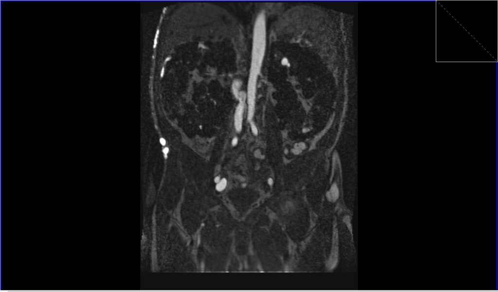
T1 FLASH 3D dynamic coronal image shows Abdominal Wall AVM
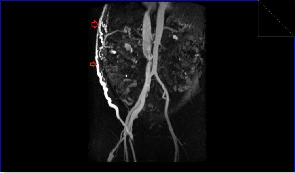
FLASH 3D MIP coronal image shows Abdominal Wall AVM
References
- Alessandrino F, Maira A, Tarantino CC. “US and MRI features in venous vascular malformation of the abdominal wall. A case report.” J Ultrasound. 2012 Sep;15(3):171–173. Published online 2012 Mar 13. doi: 10.1016/j.jus.2012.02.006. PMID: 23450707.
- Aravinda, P. S., Nag, H. H., Betigeri, V. K. M., Sakhuja, P., & Sharma, A. (2017). An Unusual Case of Giant Arterio-Venous Malformation of Anterior Abdominal Wall. Journal of Clinical Diagnosis and Research, 11(8), PD07–PD08.
- Bashir, U., Shah, S., Jeph, S., O’Keeffe, M., & Khosa, F. (2017). Magnetic Resonance (MR) Imaging of Vascular Malformations. Pol J Radiol, 82, 731-741. doi: 10.12659/PJR.903491. PMCID: PMC5894044. PMID: 29657639.
- Laakso, A., & Hernesniemi, J. (2012). Arteriovenous malformations: epidemiology and clinical presentation. Neurosurg Clin N Am, 23(1), 1-6. doi: 10.1016/j.nec.2011.09.012. PMID: 22107853.