Wrist Hemangioma MRI
A wrist hemangioma is a benign (non-cancerous) vascular tumor formed due to an abnormal collection of blood vessels. It is a type of hemangioma that specifically occurs in or around the wrist. These growths are generally painless but can cause discomfort or functional limitations depending on their size and location.
Causes of Wrist Hemangioma
The exact cause of hemangiomas is unknown. However, they are believed to result from the overgrowth of blood vessels during embryonic development. Several factors may contribute:
- Genetics: Family history may increase the likelihood of hemangiomas.
- Hormonal Influences: Rapid growth during infancy is often linked to hormonal changes.
- Congenital Factors: Hemangiomas are often present at birth or develop shortly after.
Symptoms
The symptoms of a wrist hemangioma depend on its size, depth, and location. Common symptoms include:
- Visible lump or swelling: A red, bluish, or purplish lump on or near the wrist.
- Discomfort or pain: Pain may occur due to pressure on surrounding tissues, nerves, or joints.
- Functional limitations: Restricted wrist movement or grip strength in some cases.
- Warmth over the lump: The hemangioma may feel warm to the touch due to increased blood flow.
- Cosmetic concerns: Depending on size, a hemangioma may cause aesthetic concerns.
- Skin changes: Occasionally, the skin over the hemangioma may appear stretched, discolored, or ulcerated.
Diagnosis
To diagnose a wrist hemangioma, the following methods are commonly used:
Clinical Examination:
- A physical examination of the wrist to check for the size, color, and texture of the lump.
- Assessment of pain and range of motion.
Imaging Studies:
- Ultrasound: To determine the vascular nature of the mass and its blood flow pattern.
- MRI (Magnetic Resonance Imaging): Provides detailed images of the hemangioma’s size, depth, and impact on surrounding tissues.
- CT Scan: May be used in rare cases for further evaluation.
Treatment of Wrist Hemangioma
Treatment depends on the size, location, and symptoms of the hemangioma. Options include:
Observation:
- Small, asymptomatic hemangiomas are often monitored without intervention as they may regress on their own.
Medications:
- Beta-Blockers (e.g., Propranolol): Commonly used for infantile hemangiomas to reduce growth and shrink the lesion.
- Corticosteroids: Can help reduce inflammation and slow the growth of the hemangioma.
Sclerotherapy:
- Injection of a sclerosing agent to reduce the size of the vascular lesion.
Laser Therapy:
- Effective for superficial hemangiomas to reduce redness and appearance.
Surgical Excision:
- Indicated for symptomatic, large, or complicated hemangiomas that impair wrist function or cause persistent pain.
Embolization:
- Cutting off the blood supply to the hemangioma to reduce its size and symptoms, often used for deeper lesions.
MRI Appearance of wrist Hemangioma
STIR Appearance of wrist Hemangioma
On MRI STIR (Short Tau Inversion Recovery) sequences, a wrist hemangioma appears as a hyperintense (bright) lesion due to its high water content and vascular nature. STIR effectively suppresses fat signals, enhancing the visualization of the hemangioma, especially if it has a significant soft tissue component or edema surrounding it.
PD Fat-Saturated Appearance of wrist Hemangioma
On Proton Density Fat-Saturated (PD FAT SAT) sequences, wrist hemangiomas appear hyperintense relative to muscle, as PD-weighted images are sensitive to fluid content and soft tissue details. The fat saturation further suppresses the surrounding fat signal, improving contrast and making the vascular lesion more prominent.
T1 Appearance of wrist Hemangioma
On T1-weighted MRI images, wrist hemangiomas typically appear as hypointense to isointense lesions compared to surrounding muscle and fat. Due to the low protein and fat content within the lesion, T1-weighted imaging is less sensitive for detecting hemangiomas but can help evaluate the presence of fatty components if present.
T2 Appearance of wrist Hemangioma
On T2-weighted MRI sequences, wrist hemangiomas are hyperintense (bright) due to their high fluid content and slow-flowing blood within the vascular spaces. This bright signal on T2 is characteristic of hemangiomas and helps differentiate them from other solid or fibrous lesions.
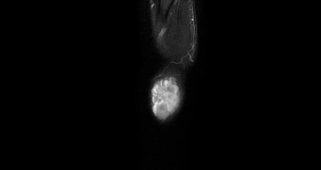
T2 GRE (T2*) Appearance of wrist Hemangioma
On T2-weighted MRI sequences, wrist hemangiomas are hyperintense (bright) due to their high fluid content and slow-flowing blood within the vascular spaces. This bright signal on T2 is characteristic of hemangiomas and helps differentiate them from other solid or fibrous lesions.
T1 Fat-Saturated Post-Contrast Appearance of wrist Hemangioma
On T1-weighted Fat-Saturated Post-Contrast images, wrist hemangiomas demonstrate intense, heterogeneous enhancement following intravenous gadolinium administration. This enhancement occurs due to the vascular nature of the lesion, where the blood vessels rapidly uptake contrast. The fat saturation helps suppress background fat signals, making the enhancement more conspicuous and allowing clear delineation of the hemangioma’s margins and internal architecture.
STIR coronal image of wrist shows Hemangioma


T1 coronal image of wrist shows Hemangioma


STIR axial image of wrist shows Hemangioma


PD FS sagittal image of wrist shows Hemangioma


T2* axial image of wrist shows Hemangioma

References
- Theumann, N.H., Bittoun, J., Goettmann, S., Le Viet, D., Chevrot, A. and Drapé, J.L., 2001. Hemangiomas of the Fingers: MR Imaging Evaluation. Radiology, 218(3). Available at: https://doi.org/10.1148/radiology.218.3.r01mr18841
- Teo, E.-L. H. J., Strouse, P. J., & Hernandez, R. J. (2000). MR Imaging Differentiation of Soft-Tissue Hemangiomas from Malignant Soft-Tissue Masses. American Journal of Roentgenology, 174(6), 1623–1628. https://doi.org/10.2214/ajr.174.6.1741623
- Hawnaur, J.M., Whitehouse, R.W., Jenkins, J.P.R., & Isherwood, I. (1990) Musculoskeletal haemangiomas: comparison of MRI with CT. Skeletal Radiology, 19, 251–258. Springer Nature. Available at: https://link.springer.com/article/10.1007/BF00191666
- lum, A.G., Gillet, R., Athlani, L., Prestat, A., Zuily, S., Wahl, D., Dautel, G. and Teixeira, P.G., 2021. CT angiography and MRI of hand vascular lesions: technical considerations and spectrum of imaging findings. Insights into Imaging, 12, Article 16. Available at: https://doi.org/10.1186/s13244-021-00969-2