Colloid cyst MRI
A colloid cyst is a noncancerous tumor typically found in the brain, more specifically in the ventricular system, which is involved in the production and management of cerebrospinal fluid. These cysts are most commonly located in the third ventricle, which can affect the flow of cerebrospinal fluid and lead to increased intracranial pressure.
Causes
The exact cause of colloid cysts is unknown. They are considered congenital, meaning they are present from birth, although they usually become symptomatic in adults. The cysts are filled with gelatinous material, which is thought to arise from the embryological remnants of the neuroepithelium or endoderm.
Symptoms
Symptoms of a colloid cyst can vary depending on its size and location but commonly include:
- Headaches, often severe and sudden
- Nausea and vomiting
- Memory disturbances
- Behavioral changes
- Confusion or disorientation
- Gait disturbances
- Drowsiness or even sudden episodes of loss of consciousness
In severe cases, due to the blockage of cerebrospinal fluid flow, a colloid cyst can lead to hydrocephalus , causing increased intracranial pressure and, potentially, life-threatening complications.
Diagnosis
Diagnosing a colloid cyst typically involves imaging studies, including:
- CT Scan (Computed Tomography): This can quickly show the presence of a cyst but might not provide detailed information.
- MRI (Magnetic Resonance Imaging): MRI is more sensitive and can provide detailed images of the brain’s ventricular system and the cyst.
In some cases, neurological evaluations and tests are conducted to assess the impact of the cyst on brain function.
Treatment
Treatment for a colloid cyst depends on the size of the cyst, its location, the symptoms it causes, and the overall health of the patient. Treatment options include:
- Observation: Small, asymptomatic cysts might simply be monitored over time with regular imaging tests to see if they grow or cause symptoms.
- Surgical removal: This is often recommended for cysts causing symptoms or showing signs of growth. Surgical approaches can vary:
- Endoscopic removal: A minimally invasive technique where tools are guided through small holes in the skull.
- Craniotomy: A more invasive procedure where part of the skull is removed to access the cyst.
In some cases, a shunt may be placed to manage hydrocephalus if it cannot be resolved by removing the cyst alone.
MRI appearance of colloid cyst
MRI T1 Appearance of Colloid Cyst
On T1-weighted MRI images, colloid cysts typically appear iso-intense to hyper-intense compared to the surrounding brain tissue. The signal intensity on T1 can vary depending on the protein content and viscosity of the cyst. Higher protein concentration tends to result in a brighter (hyper-intense) signal. The relatively homogeneous appearance on T1-weighted images helps in distinguishing colloid cysts from other types of cystic lesions within the brain.
MRI T2 Appearance of Colloid Cyst
On T2-weighted MRI images, colloid cysts can exhibit a range of signal intensities. These cysts often show a characteristic appearance with a central low T2 signal surrounded by a high peripheral T2 signal. This pattern is due to the varying contents of the cyst, with the central part possibly containing more proteinaceous material, leading to a darker (low T2) signal, while the periphery may contain less dense fluid, resulting in a brighter (high T2) signal. This distinctive feature aids in the identification and diagnosis of colloid cysts.
MRI FLAIR Appearance of Colloid Cyst
On FLAIR (Fluid-Attenuated Inversion Recovery) images, colloid cysts generally appear with a mixed signal intensity. The central part of the cyst might show a low signal, similar to its appearance on T2-weighted images, due to its dense, proteinaceous content. The peripheral part usually presents a higher signal. The FLAIR sequence is particularly useful in suppressing the signal from cerebrospinal fluid (CSF), making it easier to identify the cyst against the suppressed background of the ventricular system.
MRI DWI (b0, b1000) and ADC Appearance of Colloid Cyst
Diffusion-weighted imaging (DWI) of colloid cysts typically shows a variable signal depending on the cyst’s content. On b0 images, the cyst may appear with intermediate to high signal intensity. On b1000 images, the signal can vary; however, colloid cysts often show a relatively high signal due to restricted diffusion. The Apparent Diffusion Coefficient (ADC) map typically shows low ADC values, reflecting the restricted diffusion within the cyst. This pattern is indicative of the dense, viscous material inside the colloid cyst, which limits the movement of water molecules.
MRI T1 Post-Contrast Appearance of Colloid Cyst
On T1 post-contrast images, colloid cysts typically do not show significant enhancement. The lack of enhancement is due to the fact that these cysts are usually lined by a simple cuboidal or columnar epithelium without a significant vascular supply. However, slight peripheral enhancement may be seen in some cases due to a mild inflammatory response or the presence of a thin capsule.
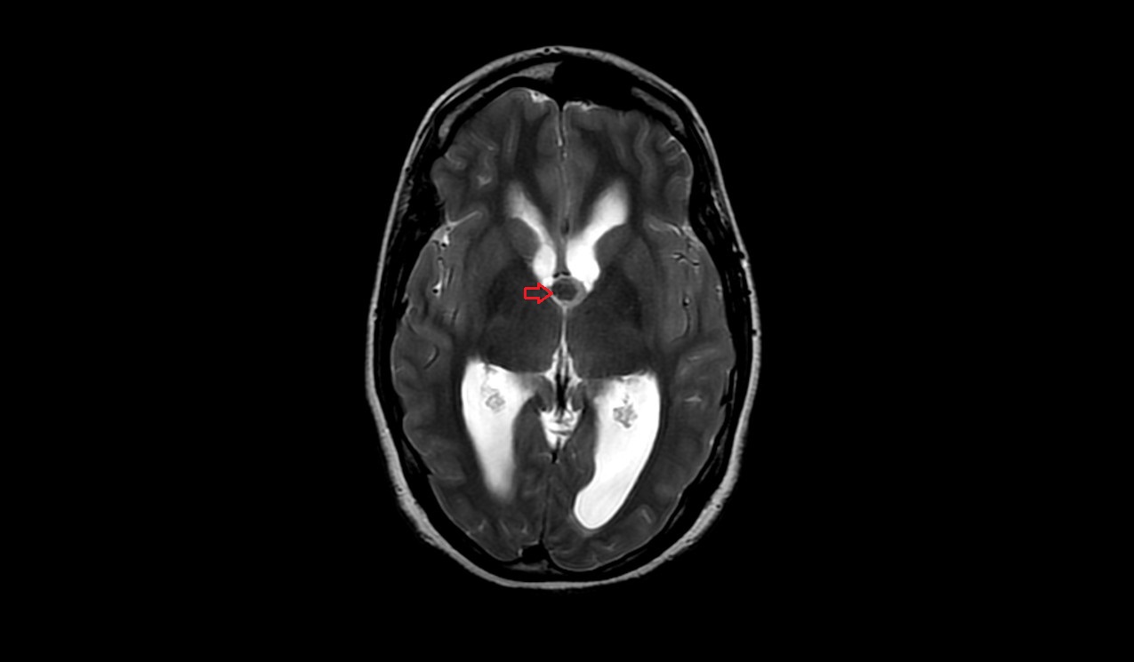
T2 axial images shows Colloid Cyst of brain

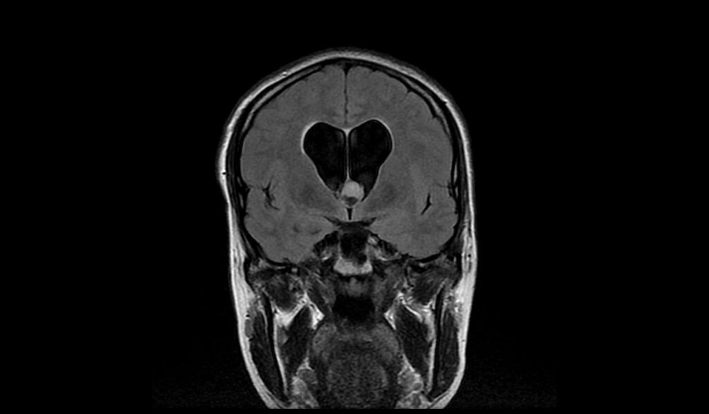
FLAIR coronal images shows Colloid Cyst of brain
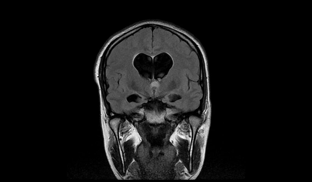
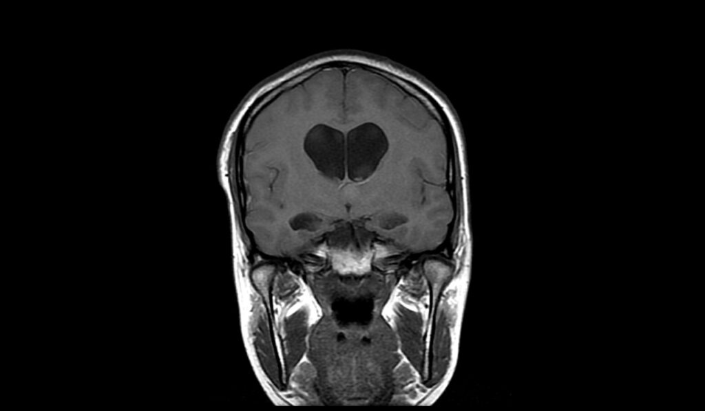
T1 coronal images shows Colloid Cyst of brain
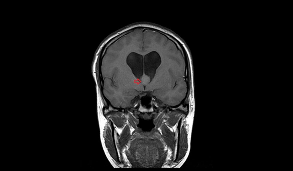
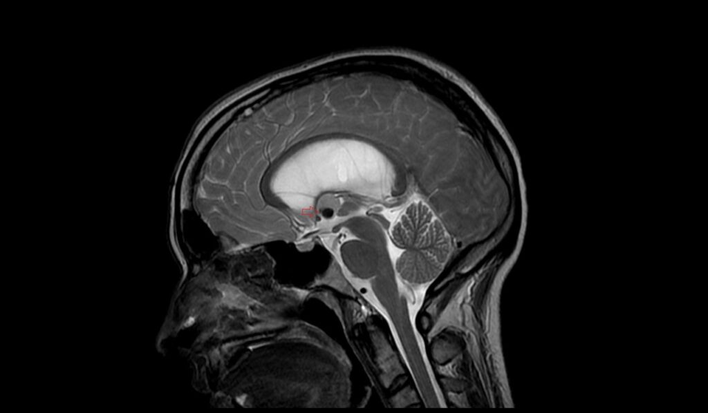
T2 sagittal images shows Colloid Cyst of brain
References
- Sener, R. N. (2007). Colloid cyst: diffusion MR imaging findings. Journal of Neuroimaging, 17(2), 181-183.
- Khanpara, S. D., Day, A. L., Bhattacharjee, M. B., Riascos, R. F., Fernelius, J. P., & Westmark, K. D. (2020). The variable appearance of third ventricular colloid cysts: Correlation with histopathology and the risk of obstructive ventriculomegaly. American Journal of Neuroradiology, 41(10), 1833-1840.
- El Khoury C, Brugières P, Decq P, Cosson-Stanescu R, Combes C, Ricolfi F, Gaston A. Colloid cysts of the third ventricle: Are MR imaging patterns predictive of difficulty with percutaneous treatment? Am J Neuroradiol. 2000;21(3):489-492.
- Hamidi, H., Faizi, F. R., Rasouly, N., & Hoshang, M. M. S. (2017). CT and MRI features of pediatric-aged colloid cysts: Report of two cases. Case Reports in Radiology. https://doi.org/10.1155/2017/2467085