Cholesteatoma MRI
Cholesteatoma is an abnormal, noncancerous growth of skin cells in the middle ear behind the eardrum. It can develop as a cyst or pouch that sheds layers of old skin, which build up inside the ear. Over time, this can increase in size and cause damage to the bones of the middle ear, the inner ear, and the surrounding structures.
Causes:
- Eustachian Tube Dysfunction: Poor function of the eustachian tube can lead to chronic ear infections, which can contribute to the formation of a cholesteatoma.
- Ear Infections: Recurrent ear infections can lead to negative pressure in the middle ear, pulling a part of the eardrum inward to form a pouch that can accumulate dead skin cells.
- Congenital: Some people are born with a small remnant of skin trapped in the middle ear that can develop into a cholesteatoma.
- Trauma: Injury or surgery to the ear can also cause a cholesteatoma.
Symptoms:
- Persistent ear drainage (otorrhea), often with a foul odor
- Hearing loss in the affected ear
- Ear pain or discomfort
- A feeling of fullness or pressure in the ear
- Dizziness or balance problems (in advanced cases)
- Facial muscle weakness (if the cholesteatoma has damaged the facial nerve)
Diagnosis
Diagnosis typically involves:
- Medical History and Physical Examination: Reviewing symptoms and performing an ear examination with an otoscope.
- Audiometry: Hearing tests to assess the extent of hearing loss.
- Imaging Tests: CT scans or MRI to determine the size and extent of the cholesteatoma and to check for any bone erosion or other complications.
Treatment
The primary treatment for cholesteatoma is surgical removal. The type of surgery depends on the size and extent of the growth:
- Microsurgical Techniques: These are used to remove the cholesteatoma while trying to preserve hearing and other functions.
- Reconstruction: Post-removal, reconstructive surgery may be necessary, especially if the bones of the middle ear are damaged.
MRI Appearance of Cholesteatoma
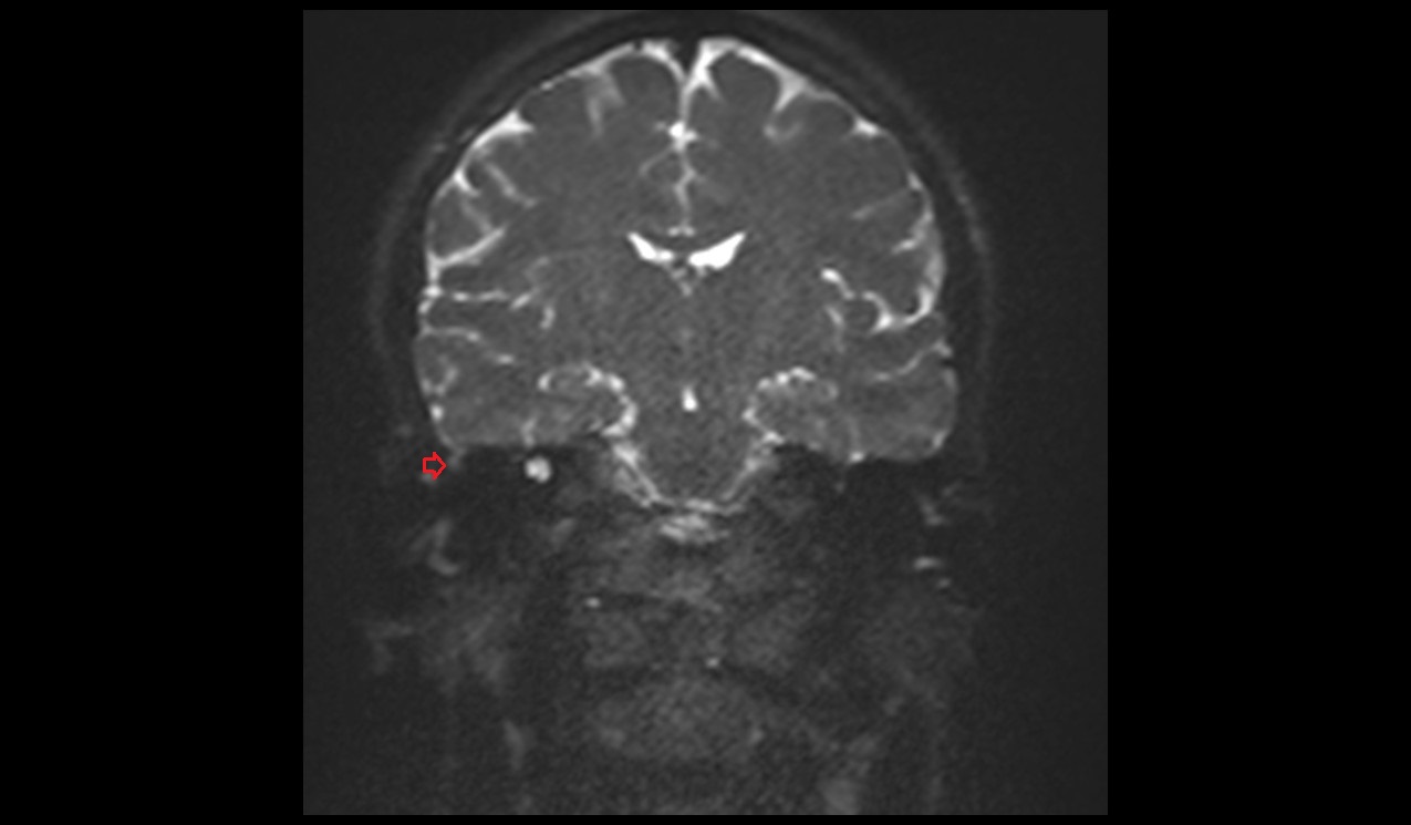
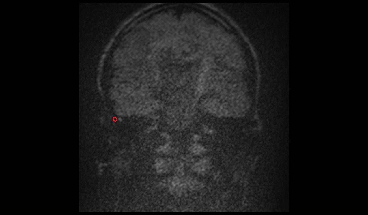
DWI b0 and b1000 Appearance of Cholesteatoma
In MRI diffusion-weighted imaging (DWI), b0 and b1000 sequences are crucial for identifying cholesteatomas. The b0 image, which involves no diffusion weighting, usually shows high signal intensity from cholesteatomas, appearing bright compared to surrounding tissue. The b1000 image, which applies a diffusion weighting factor of 1000 s/mm², helps in detecting diffusion restrictions typical of cholesteatomas. These lesions appear hyperintense on b1000 images, indicating restricted diffusion due to their keratin content and cellular debris. The comparison of signal intensities between b0 and b1000 can aid in distinguishing cholesteatomas from other lesions like cysts or inflammatory tissue.
ADC map Appearance of Cholesteatoma
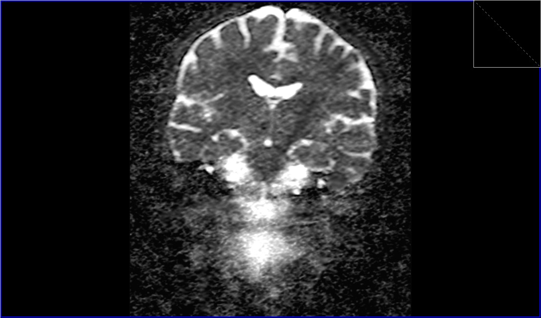
The Apparent Diffusion Coefficient (ADC) maps provide quantitative data on diffusion within tissues and are particularly useful for characterizing cholesteatomas. In cholesteatomas, ADC values are usually low, reflecting the restricted diffusion within these lesions.
T1-Weighted Appearance of Cholesteatoma
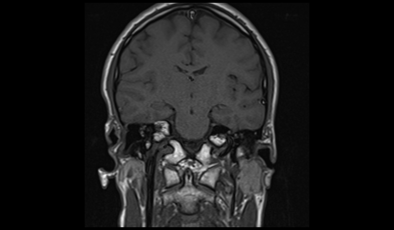
In T1-weighted MRI sequences, cholesteatomas usually appear hypointense or isointense relative to brain tissue. This imaging feature, combined with the characteristics seen in other sequences, aids in the differential diagnosis.
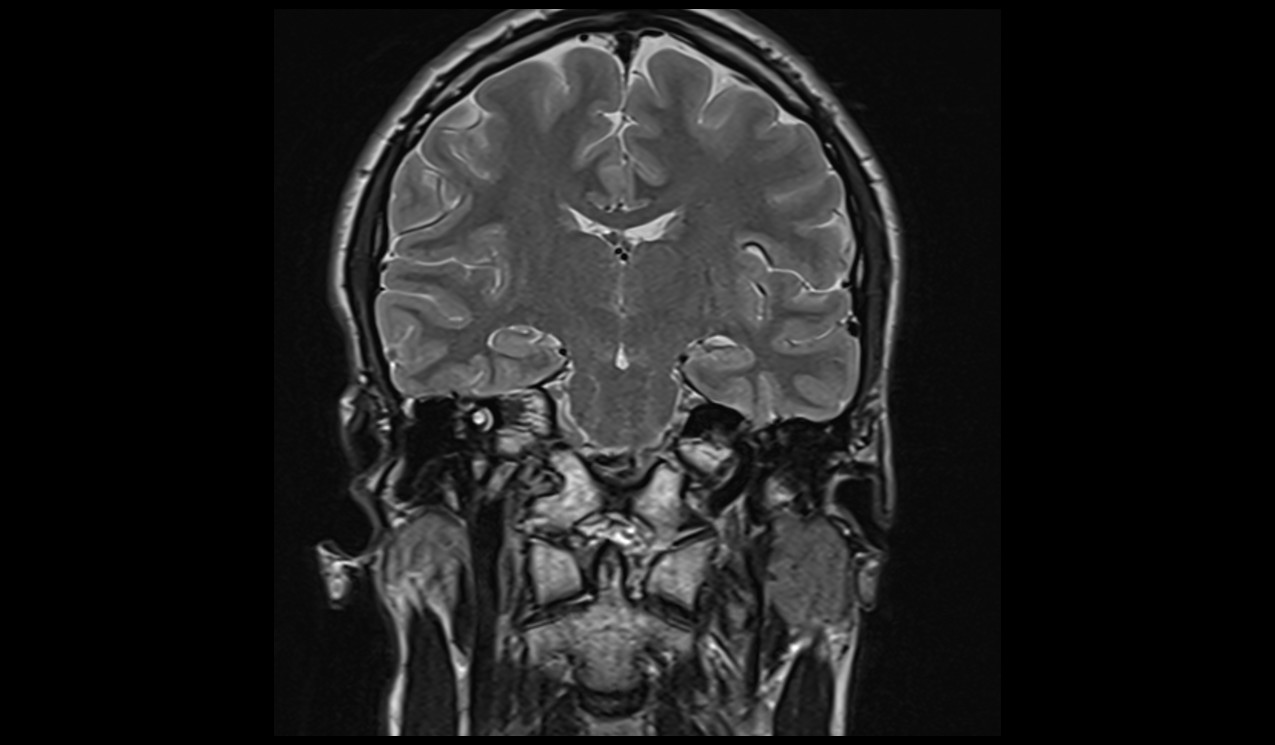
T2-Weighted Appearance of Cholesteatoma
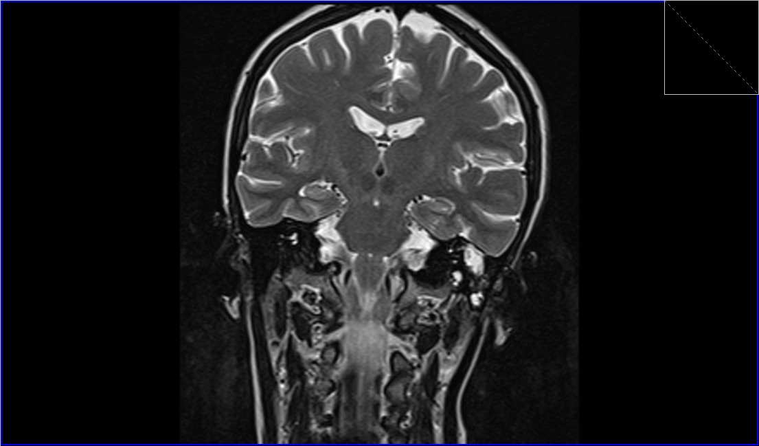
On T2-weighted MRI sequences, cholesteatomas usually appear as hyperintense masses due to their high water content.
MRI DWI b0 coronal image shows Cholesteatoma


MRI DWI b1000 coronal image shows Cholesteatoma


MRI T2 coronal image shows Cholesteatoma
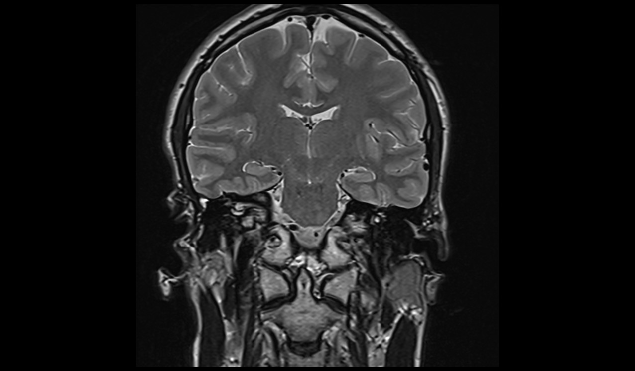

MRI T1 coronal image shows Cholesteatoma


Case Study 2
MRI DWI b100 coronal image shows Cholesteatoma
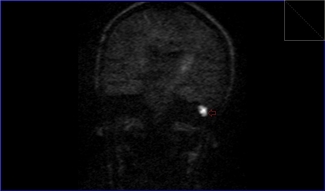
MRI DWI b0 coronal image shows Cholesteatoma

MRI DWI ADC coronal image shows Cholesteatoma
MRI T2 coronal image shows Cholesteatoma
References
- Fitzek, C., Mewes, T., Fitzek, S., Mentzel, H.-J., Hunsche, S., & Stoeter, P. (2002). Diffusion-weighted MRI of cholesteatomas of the petrous bone. Journal of Magnetic Resonance Imaging, 15(6), 636-641. https://doi.org/10.1002/jmri.10118
- Kumar, J. (2024). Diffusion-weighted magnetic resonance imaging of cholesteatoma: Navigating the multifarious techniques. Indian Journal of Radiology and Imaging, 34(1), 3–5. https://doi.org/10.1055/s-0043-1777292
- Vaid, S., Kamble, Y., Vaid, N., Bhatti, S., Rawat, S., Nanivadekar, A., & Karmarkar, S. (2013). Role of magnetic resonance imaging in cholesteatoma: The Indian experience. Indian Journal of Otolaryngology and Head & Neck Surgery, 65(Suppl 3), 485-492. https://doi.org/10.1007/s12070-011-0360-1
- Aikele, P., Kittner, T., Offergeld, C., Kaftan, H., Hüttenbrink, K.-B., & Laniado, M. (2003). Diffusion-weighted MR imaging of cholesteatoma in pediatric and adult patients who have undergone middle ear surgery. American Journal of Roentgenology, 181(1). https://doi.org/10.2214/ajr.181.1.1810261


