Compressed Sensing (CS) \ Hyper Sense MRI
Compressed Sensing
Compressed Sensing (CS) MRI is a groundbreaking technique that aims to accelerate the acquisition of MRI data while maintaining image quality. Traditional MRI methods require collecting a large amount of raw data to reconstruct high-resolution images. CS takes advantage of the fact that MRI scan images can be represented using fewer data points in certain domains without a significant loss of information.
How Compressed Sensing MRI Works
Transform Sparsity:
Many MRI images are not sparse in their native spatial domain, but they become sparse when transformed into domains such as wavelet, Fourier, or total variation. This means that most of the signal energy is concentrated in a few significant coefficients, while the rest are near zero or negligible. This principle is referred to as transform sparsity and is essential for enabling compressed sensing.
Pseudo-Random Undersampling:
In conventional MRI, k-space is sampled in a uniform grid. CS MRI uses pseudo-random or variable-density sampling patterns, particularly undersampling the high-frequency components while preserving central (low-frequency) k-space regions. These patterns introduce incoherent artifacts, which appear as noise and can be removed during reconstruction, avoiding structured aliasing.
Nonlinear Image Reconstruction:
Reconstruction in CS MRI involves solving an optimization problem that seeks the sparsest possible image consistent with the acquired data. This is typically formulated using:
L1 norm minimization to promote sparsity,
L2 norm minimization to ensure data fidelity.
The optimization process is performed using iterative algorithms such as nonlinear conjugate gradient, iterative soft thresholding, or other sparsity-promoting solvers.
CS can also be combined with parallel imaging (PI) methods to enhance acceleration even further.
The benefits of Compressed Sensing MRI are significant. By acquiring fewer data points, CS MRI substantially reduces scan time. This acceleration is particularly valuable for capturing dynamic processes or imaging patients who struggle to remain still during longer scans.
Compressed Sensing has transformed various fields of medical imaging:
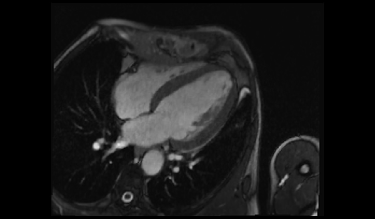
Compressed Sensing (CS) FB cine imaging of the heart

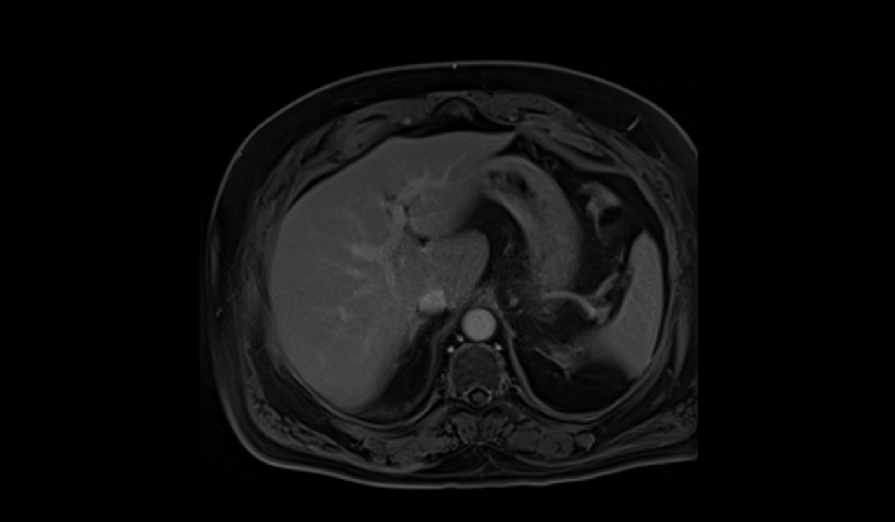
Compressed Sensing (CS) FB GRASP-VIBE imaging of the liver

Benefits of Compressed Sensing (CS) in Cardiac Imaging:
In the realm of cardiac imaging, the introduction of CS Cardiac Cine stands out as a remarkable innovation. This pioneering approach has significantly reduced the time required for a Cardiac Cine scan, which previously took approximately six minutes involving multiple instances of breath-holding. However, with the integration of Compressed Sensing, this scan can now be completed within an astonishingly short span of 15-25 seconds, all while allowing patients to breathe naturally throughout the procedure. This not only enhances patient comfort but also ensures precise quantification by capturing the entire cardiac cycle.
T2 TRUEFISP CINE performed on an uncooperative patient.

scan acquisition time 10sec.
CS free breathing cine performed in an uncooperative patient.

scan acquisition time 2sec.
Benefits of CS in Abdominal Imaging:
Similarly, the adoption of Compressed Sensing GRASP-VIBE has brought about transformative advantages in abdominal MRI imaging. In contrast to the traditional approach that demanded intricate timing for contrast administration, repeated breath-holding, and patient cooperation, CS GRASP-VIBE captures all essential data in a single continuous session under unrestricted breathing conditions. This eliminates the complexities stemming from timing constraints and respiratory artifacts. Moreover, Compressed Sensing empowers high-resolution 3D imaging of the pancreatic and biliary duct system within a brief breath-hold or just 1-2 minutes of unhindered breathing, thanks to acceleration factors of up to 20 times.
VIBE DIXON performed on an uncooperative patient.

scan acquisition time 18sec.
CS-FB GRASP VIBE performed on an uncooperative patient.

scan acquisition time 2min.
Benefits of CS in Musculoskeletal (MSK) and Vascular Imaging:
The impact of Compressed Sensing technology extends further to neurology and musculoskeletal imaging. Applications like CS SPACE, CS SEMAC, and CS TOF have facilitated isotropic 3D imaging of the brain and MSK joints with heightened clinical significance. Typically, these scans required 5-6 minutes, often resulting in compromises in spatial resolution to expedite scan times. However, with Compressed Sensing, 3D brain scans featuring 1mm isotropic resolution can be achieved in only 3 minutes per scan, and high-resolution MSK imaging becomes achievable with shorter scan durations. Notably, CS SEMAC empowers high-quality imaging even in the presence of metal implants, leading to scan time reductions of up to 55%.
CS 3D spine with acceleration factor 6

Scan time : 3minutes
CS 3D spine with acceleration factor 11

Scan time : 2minutes
Drawbacks of CS Imaging
While Compressed Sensing (CS) MRI offers significant advantages in terms of accelerated imaging and reduced scan times, there are a few disadvantages associated with this technique. Here are some of the key disadvantages of CS MRI:
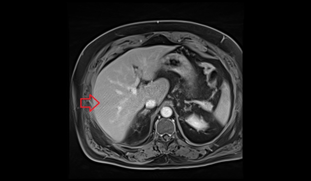
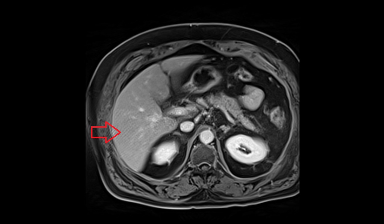
Image Artifacts: CS MRI can be more sensitive to certain types of artifacts, such as noise and aliasing artifacts, due to the undersampling of k-space. These artifacts can affect image quality and diagnostic accuracy.
Compressed Sensing (CS) artifacts in liver imaging.


Loss of Image Quality: Although CS MRI aims to maintain image quality, there can be a trade-off between scan time reduction and image fidelity. In some cases, particularly with high acceleration factors, there may be a compromise in image resolution or signal-to-noise ratio
CS acceleration factor 6

CS ACCELERATION FACTOR 16

References
- Lustig, M., Donoho, D. L., Santos, J. M., & Pauly, J. M. (2007). Compressed sensing MRI. IEEE Signal Processing Magazine, 25(2), 72-82.
- Lustig, M., Donoho, D. L., & Pauly, J. M. (2008). Sparse MRI: The application of compressed sensing for rapid MR imaging. Magnetic Resonance in Medicine, 58(6), 1182-1195.
- Otazo, R., Kim, D., & Sodickson, D. K. (2015). Compressed sensing MRI: A look back and forward. Journal of Magnetic Resonance Imaging, 41(1), 1-12.
- Lustig, M., & Pauly, J. M. (2010). SPIRiT: Iterative self-consistent parallel imaging reconstruction from arbitrary k-space. Magnetic Resonance in Medicine, 64(2), 457-471.
- European Society of Radiology. (2016). Philips advances digital MR with next generation Ingenia MR-RT platform for personalized therapy planning. Retrieved from https://www.myesr.org/article/5232
- Siemens Healthineers. (2021). Compressed Sensing Cardiac Cine. Retrieved from https://www.siemens-healthineers.com/magnetic-resonance-imaging/options-and-upgrades/compressed-sensing-cardiac-cine
- Philips Healthcare. (2021). Philips MR Acceleration technology. Retrieved from https://www.usa.philips.com/healthcare/solutions/magnetic-resonance/compressed-sense


